


 下载产品说明书
 用小程序,查商品更便捷


 收藏
收藏
 对比
对比 咨询
咨询
Specificity/Sensitivity
参考图片
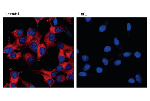
Confocal immunofluorescent analysis of HeLa cells, untreated (left), or TNF-α-treated (#8902, 10 ng/ml for 15 min, right) using IκBα (L35A5) Mouse mAb (Amino-terminal Antigen) (red). Blue pseudocolor = DRAQ5® #4084 (fluorescent DNA dye).
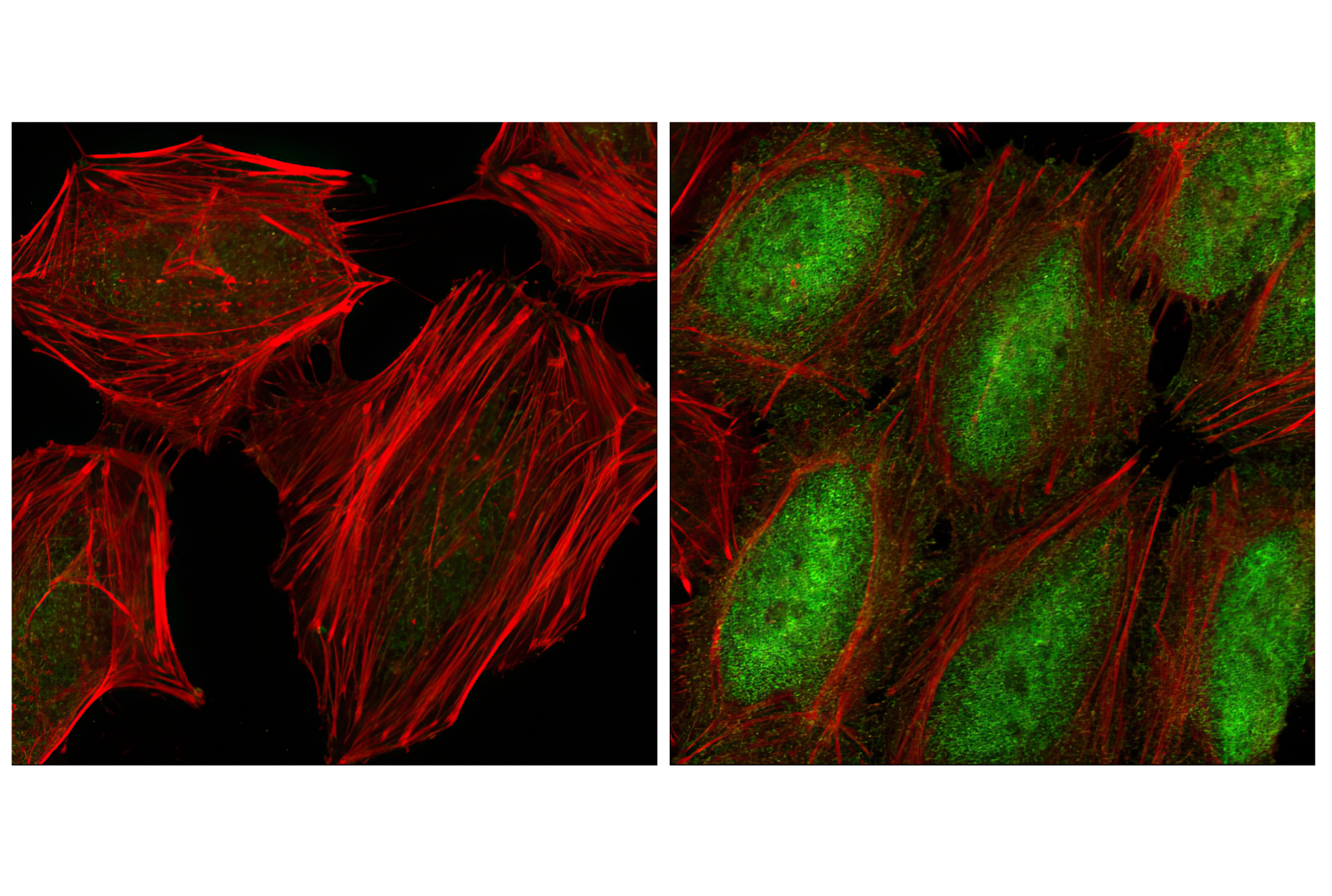
Confocal immunofluorescent analysis of HeLa cells, untreated (left) and TNF-α treated (#8902 at 20 ng/ml for 20 min, right), using Phospho-NF-κB p65 (Ser536) (93H1) Rabbit mAb (green). Actin filaments have been labeled with Alexa Fluor® phalloidin 555 (red).
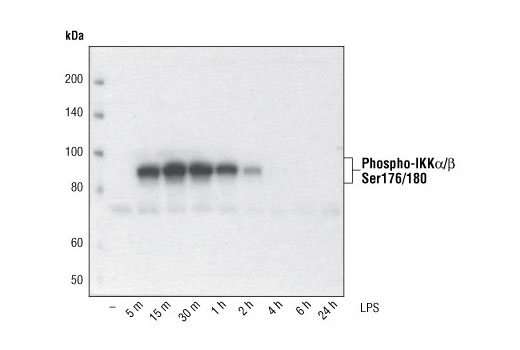
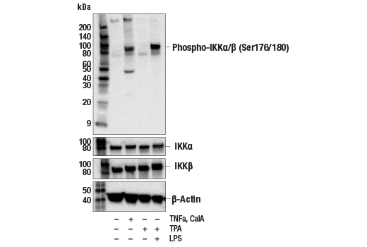
Western blot analysis of extracts from THP-1 cells, differentiated with TPA (#9905, 80 nM for 24h) and treated with 1 μg/ml LPS for the indicated times, using Phospho-IKKα/β (Ser176/180) (16A6) Rabbit mAb.
Western blot analysis of extracts from THP-1 cells, differentiated with TPA (#9905, 80 nM for 24h) and treated with 1 μg/ml LPS for the indicated times, using Phospho-NF-κB p65 (Ser536) (93H1) Rabbit mAb (upper) and NF-κB p65 (C22B4) Rabbit mAb #4764 (lower).
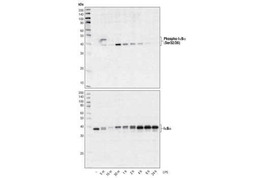
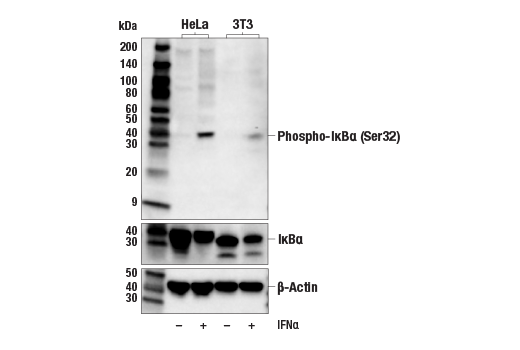
Western blot analysis of extracts from THP-1 cells, differentiated with TPA (#9905, 80 nM for 24h) and treated with 1 μg/ml LPS for the indicated times, using Phospho-IκBα (Ser32/36) (5A5) Mouse mAb #9246 (upper) and IκBα (L35A5) Mouse mAb (Amino-terminal Antigen) (lower).
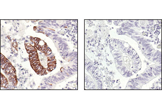
Immunohistochemical analysis of paraffin-embedded human colon carcinoma, untreated (left) or λ phosphatase-treated (right), using Phospho-IKKα/β (Ser176/180) (16A6) Rabbit mAb.
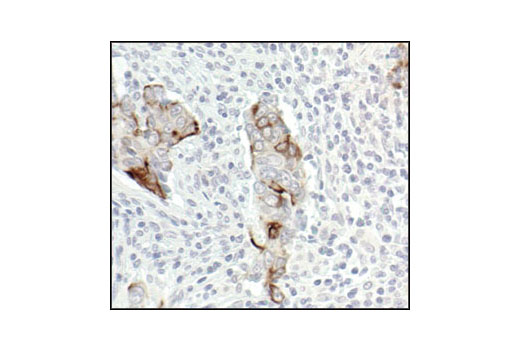
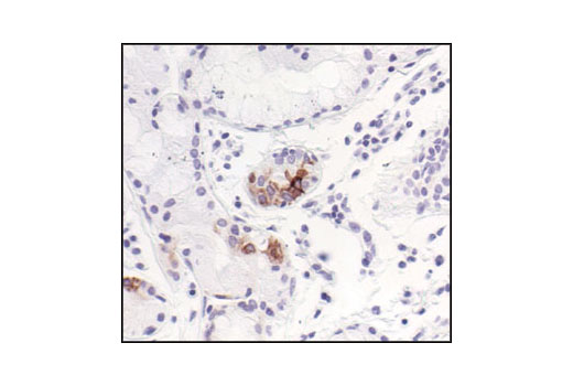
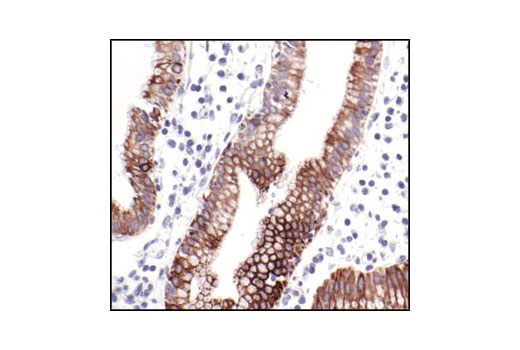
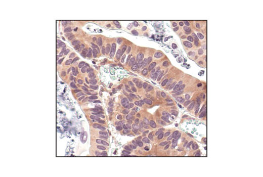
Immunohistochemical analysis of paraffin-embedded human gall bladder (chronic cholecystitis), using Phospho-IKKα/β (Ser176/180) (16A6) Rabbit mAb.
Immunohistochemical analysis of paraffin-embedded human lung (chronic bronchitis), using Phospho-IKKα/β (Ser176/180) (16A6) Rabbit mAb.
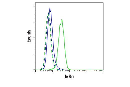
Flow cytometric analysis of HeLa cells, using IκBα (L35A5) Mouse mAb (Amino-terminal Antigen) (blue) compared to a nonspecifc negative control antibody (red).
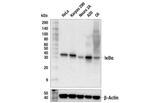
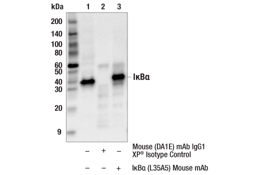
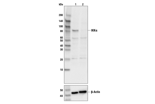
Western blot analysis of extracts from HeLa, NIH/3T3 and PC12 cells, using IkBα (L35A5) Mouse mAb (Amino-terminal Antigen).
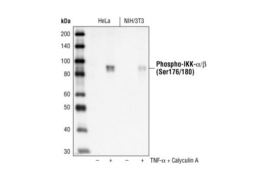
Western blot analysis of extracts from TNF-alpha and Calyculin A treated HeLa and NIH/3T3 cells, using Phospho-IKKα/β (Ser176/180) (16A6) Rabbit mAb.
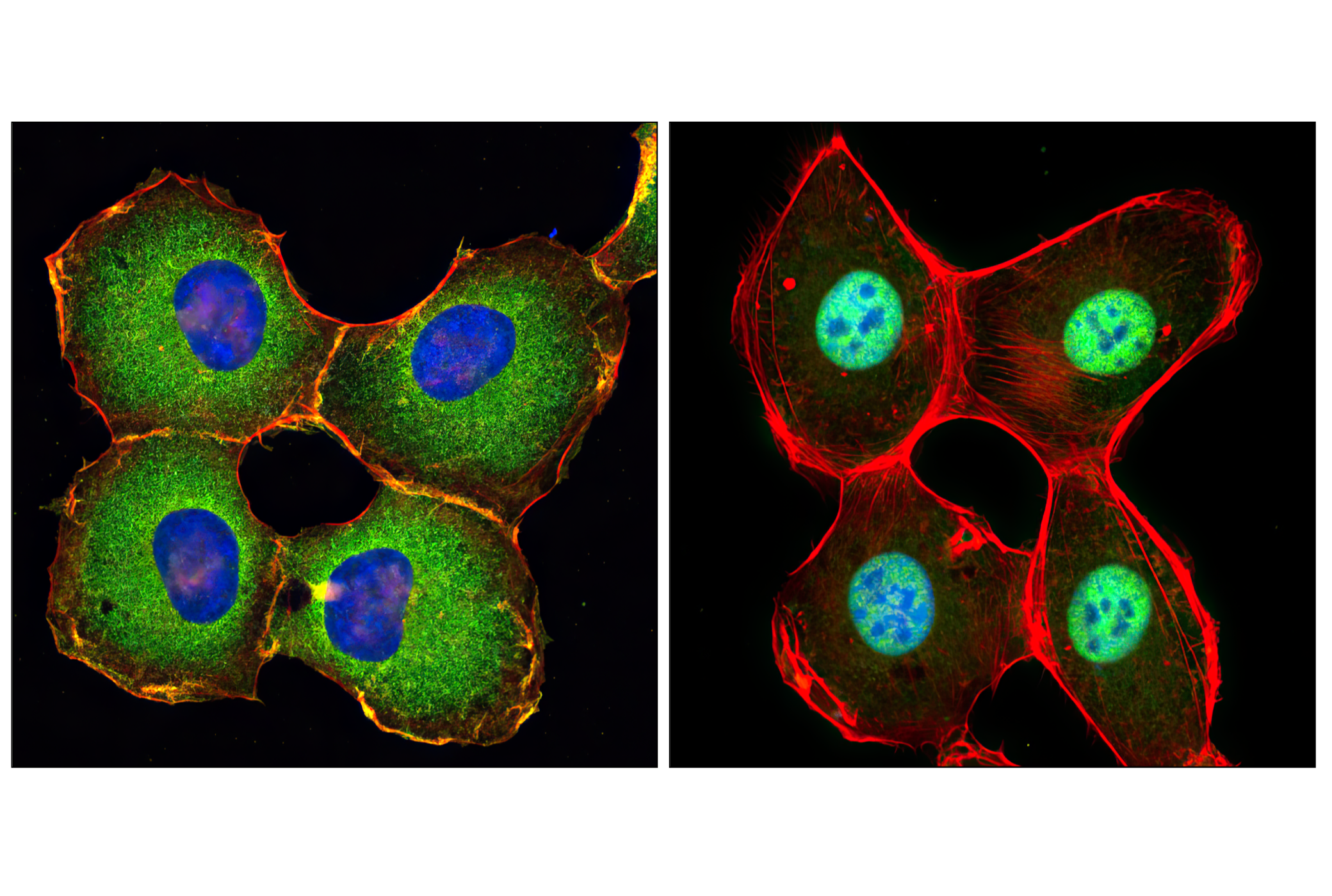
Confocal immunofluorescent analysis of HT-1080 cells, untreated (left) or treated with hTNF-α #8902 (20 ng/ml, 20 min) (right), using NF-κB p65 (D14E12) XP® Rabbit mAb (green). Actin filaments were labeled with DY-554 phalloidin (red). Blue pseudocolor = DRAQ5® #4084 (fluorescent DNA dye).
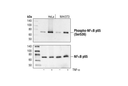
Western blot analysis of extracts from HeLa and NIH/3T3 cells, untreated or TNF-α treated (#2169, 20 ng/ml for 5 minutes), using Phospho-NF-κB p65 (Ser536) (93H1) Rabbit mAb (upper) or NF-κB p65 Antibody #3034 (lower).
Immunohistochemical analysis of paraffin-embedded human lung carcinoma, using IκBα (L35A5) Mouse mAb (Amino-terminal Antigen).
Immunohistochemical analysis of paraffin-embedded human renal adenocarcinoma, using IκBα (L35A5) Mouse mAb (Amino-terminal Antigen).
Immunohistochemical analysis of paraffin-embedded human leiomyoma, using IκBα (L35A5) Mouse mAb (Amino-terminal Antigen).
Immunohistochemical analysis of paraffin-embedded human colon carcinoma, showing cytoplasmic localization, using Phospho-IKKα/β (Ser176/180) (16A6) Rabbit mAb.
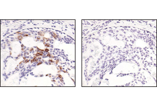
Immunohistochemical analysis of paraffin-embedded human breast carcinoma, using Phospho-IKKα/β (Ser176/180) (16A6) Rabbit mAb in the presence of control peptide (left) or Phospho-IKK-alpha/beta (Ser176/180) Blocking Peptide #1023 (right).
Western blot analysis of extracts from HeLa and NIH/3T3 cells, untreated or treated with TNF-α (#2169, 20 ng/ml) for 5 minutes, using Phospho-IκBα (Ser32) (14D4) Rabbit mAb (upper), or IκBα (44D4) Rabbit mAb #4812 (lower).
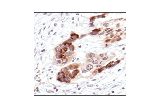
Immunohistochemical analysis of frozen H1650 xenograft, showing cytoplasmic localization using Phospho-IKKα/β (Ser176/180)(16A6) Rabbit mAb.
After the primary antibody is bound to the target protein, a complex with HRP-linked secondary antibody is formed. The LumiGLO* is added and emits light during enzyme catalyzed decomposition.
After the primary antibody is bound to the target protein, a complex with HRP-linked secondary antibody is formed. The LumiGLO* is added and emits light during enzyme catalyzed decomposition.
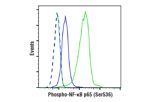
Flow cytometric analysis of HeLa cells, untreated (blue) or TNF-α-treated (green), using Phospho-NF-κB p65 (Ser536) (93H1) Rabbit mAb compared to a nonspecific negative control antibody (red).
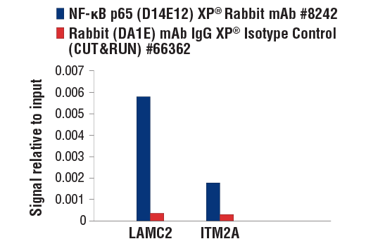
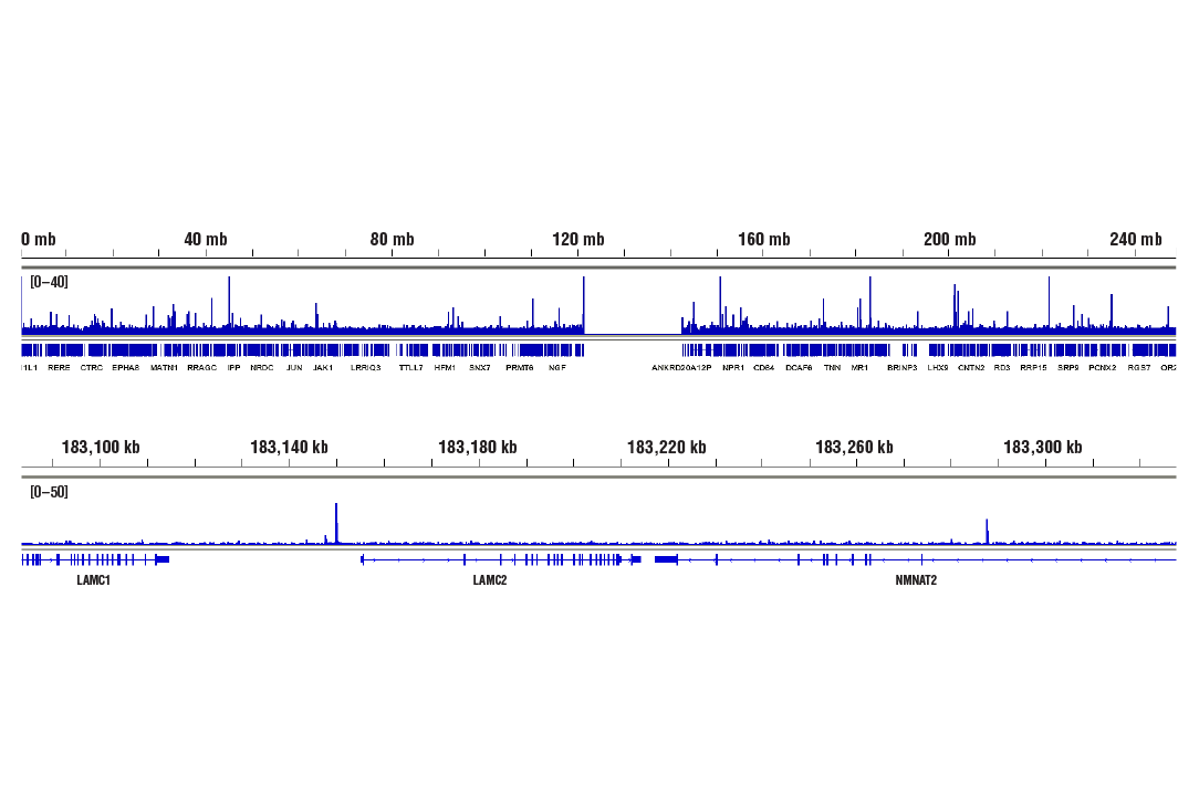
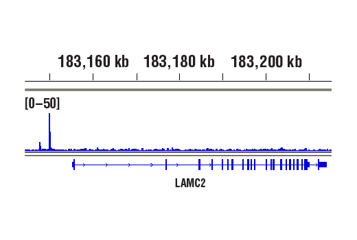
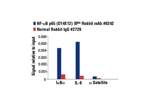
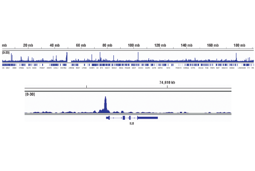
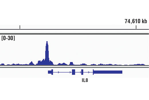
Chromatin immunoprecipitations were performed with cross-linked chromatin from 4 x 10⁶ HeLa cells treated with hTNF-α #8902 (30 ng/ml, 1 hr) and either 5 μl of NF-κB p65 (D14E12) XP® Rabbit mAb or 2 μl of Normal Rabbit IgG #2729 using SimpleChIP® Enzymatic Chromatin IP Kit (Magnetic Beads) #9003. The enriched DNA was quantified by Real-Time PCR using SimpleChIP® Human IκBα Promoter Primers #5552, human IL-8 promoter primers, and SimpleChIP® Human α Satellite Repeat Primers #4486. The amount of immunoprecipitated DNA in each sample is represented as signal relative to the total amount of input chromatin, which is equivalent to one.
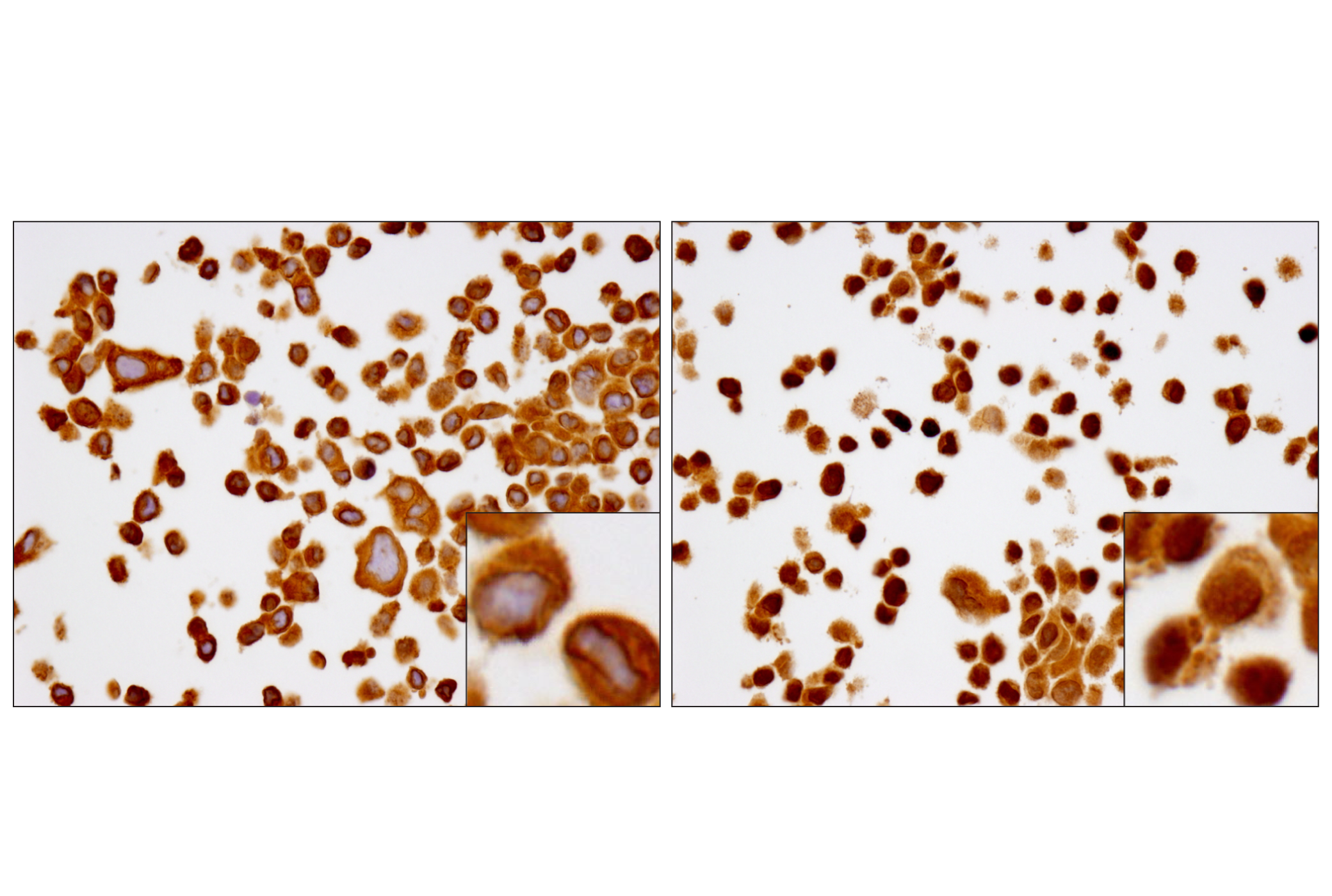
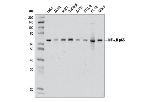
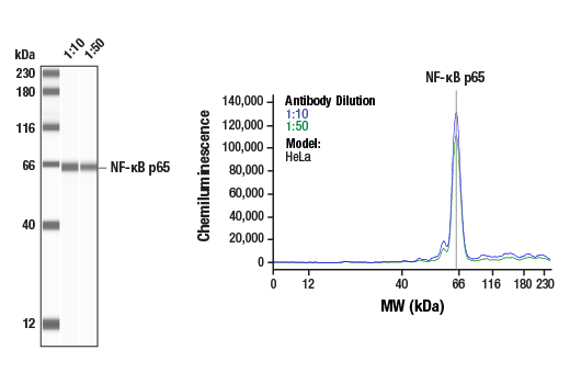
Western blot analysis of extracts from various cell lines using NF-κB p65 (D14E12) XP® Rabbit mAb.
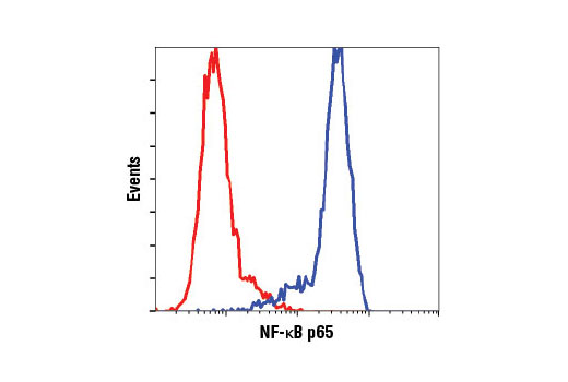
Flow cytometric analysis of HeLa cells using NF-κB p65 (D14E12) XP® Rabbit mAb (blue) compared to concentration matched Rabbit (DA1E) mAb IgG XP® Isotype Control #3900 (red).
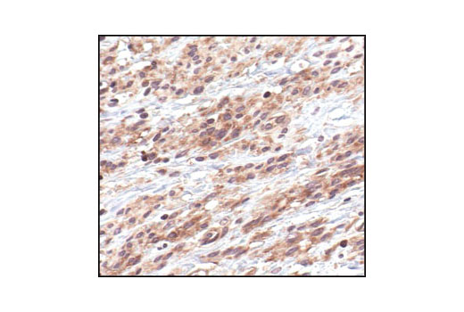
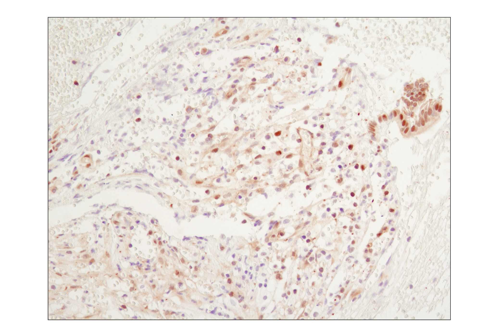
Immunohistochemical analysis of paraffin-embedded human chronic cholecystitis using NF-κB p65 (D14E12) XP® Rabbit mAb.
Western blot analysis of extracts from various cell lines using NF-κB p65 (D14E12) XP® Rabbit mAb #8242.
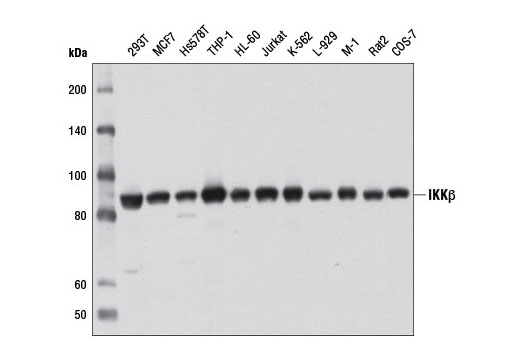
Western blot analysis of extracts from various cell lines using IKKβ (D30C6) Rabbit mAb.
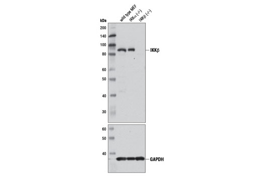
Western blot analysis of extracts from wild-type, IKKα (-/-), and IKKβ (-/-) mouse embryonic fibroblasts (MEFs) using IKKβ (D30C6) Rabbit mAb (upper) and GAPDH (14C10) Rabbit mAb #2118 (lower).
Western blot analysis of extracts from NIH/3T3, HeLa and PC12 cells, using IKKα Antibody #2682.
Western blot analysis of extracts from HeLa, NIH/3T3 and PC-12 cells using IIKKβ (2C8) Rabbit mAb #2370.
Western blot analysis of extracts from TNF-alpha and Calyculin A treated HeLa and NIH/3T3 cells, using Phospho-IKKα/β (Ser176/180) (16A6) Rabbit mAb #2697.
Western blot analysis of extracts from HeLa and NIH/3T3 cells, untreated or treated with TNF-α (#2169, 20 ng/ml) for 5 minutes, using Phospho-IκB-α (Ser32) (14D4) Rabbit mAb #2859 (upper), or IκBα (44D4) Rabbit mAb #4812 (lower).
Western blot analysis of extracts from HeLa and NIH/3T3 cells, untreated or TNF-α treated (#2169, 20 ng/ml for 5 minutes), using Phospho-NF-κB p65 (Ser536) (93H1) Rabbit mAb #3033 (upper) or NF-κB p65 Antibody #3034 (lower).
Western blot analysis of extracts from HeLa, NIH/3T3 and PC12 cells, using IκB-α (L35A5) Mouse mAb (Amino-terminal Antigen) #4814.
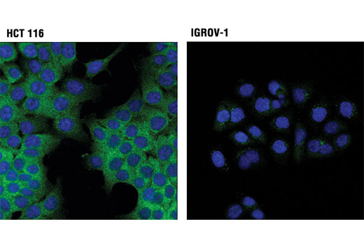
Confocal immunofluorescent analysis of HCT 116 (high expression; left) and IGROV-1 (low expression; right) cells using IKKα (3G12) Mouse mAb (green). Blue pseudocolor = DRAQ5® #4084 (fluorescent DNA dye).
Western blot analysis of extracts from various cell lines using IKKα (3G12) Mouse mAb (upper) or β-Actin (D6A8) Rabbit mAb #8457 (lower).
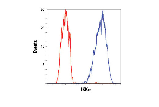
Flow cytometric analysis of HCT 116 cells using IKKα (3G12) Mouse mAb (blue) compared to concentration-matched Mouse (G3A1) mAb IgG1 Isotype Control #5415 (red). Anti-mouse IgG (H+L), F(ab')2 Fragment (Alexa Fluor® 488 Conjugate) #4408 was used as a secondary antibody.
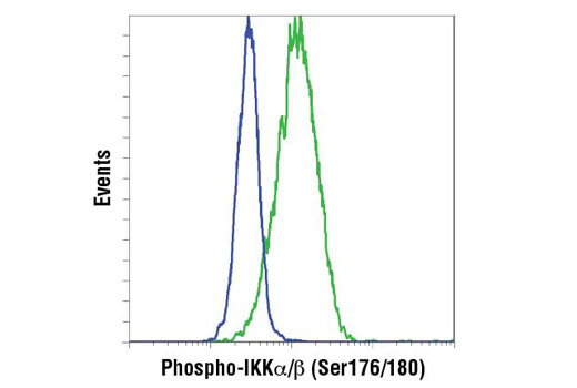
Flow cytometric analysis of THP-1 cells, untreated (blue) and with TPA and LPS (green) using IKK-α (Ser176/Ser180) phosphate Rabbit mAb. Anti-rabbit IgG (H+L), F(ab')2 Fragment (PE Conjugate) #8885 was used as a secondary antibody.

 危险品化学品经营许可证(不带存储) 许可证编号:沪(杨)应急管危经许[2022]202944(QY)
  营业执照(三证合一)