


 下载产品说明书 下载SDS
 用小程序,查商品更便捷


 收藏
收藏
 对比
对比 咨询
咨询
Product Usage Information
For optimal ChIP results, use 10 μl of antibody and 10 μg of chromatin (approximately 4 x 10⁶ cells) per IP.This antibody has been validated using SimpleChIP® Enzymatic Chromatin IP Kits.
| Application | Dilution |
|---|---|
| Western Blotting | 1:1000 |
| Immunoprecipitation | 1:100 |
| Immunohistochemistry (Paraffin) | 1:200 - 1:800 |
| Immunofluorescence (Immunocytochemistry) | 1:400 - 1:1600 |
| Flow Cytometry (Fixed/Permeabilized) | 1:200 - 1:800 |
| Chromatin IP | 1:50 |
Specificity/Sensitivity
Species Reactivity:
Human, Mouse, Rat, Hamster, Monkey, Mink, Bovine, Dog, Pig
参考图片
Flow cytometric analysis of HeLa cells using NF-κB p65 (L8F6) Mouse mAb (blue) compared to a nonspecific negative control antibody (red).流式细胞仪研究HeLa细胞,所用抗体为NF-κB p65 (L8F6) Mouse mAb (蓝色) 与非特异性的阴性对照抗体(红色)。
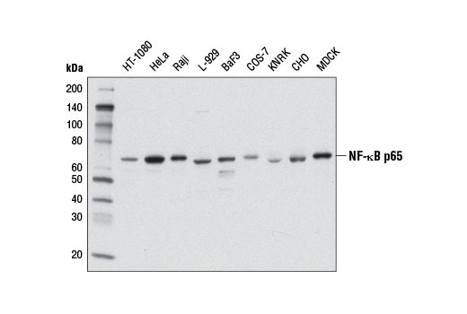
Western blot analysis of extracts from various cell lines using NF-κB p65 (L8F6) Mouse mAb.Western免疫印迹。用NF-κB p65 (L8F6) Mouse mAb研究各种类型细胞的细胞提取液。
Chromatin immunoprecipitations were performed with cross-linked chromatin from 4 x 10⁶ HeLa cells treated with Human Tumor Necrosis Factor-α (hTNF-α) #8902 (30 ng/ml, 1 hr) and either 10 μl of NF-κB p65 (L8F6) Mouse mAb or 2 μl of Normal Rabbit IgG #2729 using SimpleChIP® Enzymatic Chromatin IP Kit (Magnetic Beads) #9003. The enriched DNA was quantified by real-time PCR using SimpleChIP® Human IκBα Promoter Primers #5552, human IL-8 promoter primers, and SimpleChIP® Human α Satellite Repeat Primers #4486. The amount of immunoprecipitated DNA in each sample is represented as signal relative to the total amount of input chromatin, which is equivalent to one.染色质免疫共沉淀。HeLa细胞培养至4 x 10⁶,并用hTNF-α #8902 (30 ng/ml, 1 hr)处理后,将染色质交联到玻片上,然后用SimpleChIP® Enzymatic Chromatin IP Kit (Magnetic Beads) #9003进行免疫沉淀实验,本实验中用10 μl NF-κB p65 (L8F6) Mouse mAb 抗体或2 μl Normal Rabbit IgG #2729 。对富集的DNA做real-time PCR,所用引物为SimpleChIP® Human IκBα Promoter Primers #5552, human IL-8 promoter primers和SimpleChIP® Human α Satellite Repeat Primers #4486。每个样本中沉淀的DNA量定义为相对信号与输入的总染色质相比的数值。
Immunohistochemical analysis of paraffin-embedded HeLa cell pellets, untreated (left) or treated with Human Tumor Necrosis Factor-α (hTNF-α) #8902 (right), using NF-κB p65 (L8F6) Mouse mAb.免疫组织化学染色分析石蜡包埋HeLa细胞沉淀。未经处理(左图) 或经hTNF-α #8902 (右图)处理, 所用抗体为NF-κB p65 (L8F6) Mouse mAb。
Immunohistochemical analysis of paraffin-embedded OVCAR8 cell pellets treated with Human Tumor Necrosis Factor-α (hTNF-α) #8902 (left) or treated with SignalSilence® NF-κB p65 siRNA I #6261 (right), using NF-κB p65 (L8F6) Mouse mAb.免疫组织化学染色分析石蜡包埋OVCAR8细胞沉淀。经hTNF-α #8902 处理(左图) 或经SignalSilence® NF-κB p65 siRNA I #6261 (右图)处理, 所用抗体为NF-κB p65 (L8F6) Mouse mAb。
Immunohistochemical analysis of human chronic cholecystitis tissue using NF-κB p65 (L8F6) Mouse mAb.免疫组织化学染色分析石蜡包埋人慢性胆囊炎组织。所用抗体为NF-κB p65 (L8F6) Mouse mAb。
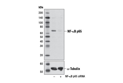
Western blot analysis of extracts from HeLa cells, transfected with 100 nM SignalSilence® Control siRNA (Unconjugated) #6568 (-) or SignalSilence® NF-κB p65 siRNA I #6261 (+), using NF-κB p65 (L8F6) Mouse mAb (upper) or α-Tubulin (11Η10) Rabbit mAb #2125 (lower). The NF-κB p65 (L8F6) Mouse mAb confirms silencing of NF-κB p65 expression, while the α-Tubulin (11Η10) Rabbit mAb is used as a loading control.Western免疫印迹。用NF-κB p65 (L8F6) Mouse mAb (上图) 或α-Tubulin (11Η10) Rabbit mAb #2125 (下图) 研究转染了100 nM SignalSilence® Control siRNA (Unconjugated) #6568 (-) 或SignalSilence® NF-κB p65 siRNA I #6261 (+)的 HeLa 细胞的细胞提取液。NF-κB p65 (L8F6) Mouse mAb 确定对NF-κB p65蛋白的沉默表达, 而 α-Tubulin (11Η10) Rabbit mAb 作为上样量的对照。
Confocal immunofluorescent analysis of HeLa cells, untreated (left) or treated with Human Tumor Necrosis Factor-α (hTNF-α) #8902 (20 ng/mL, 20 min; right), using NF-κB p65 (L8F6) Mouse mAb (green). Actin filaments were labeled with DY-554 phalloidin (red). Blue pseudocolor = DRAQ5® #4084 (fluorescent DNA dye).共聚焦免疫荧光分析HeLa细胞,未经处理 (左图)或 经hTNF-α #8902 (20 ng/ml, 20 min) (右图)处理, 所用抗体为NF-κB p65 (L8F6) Mouse mAb (绿色) 肌动蛋白微丝用DY-554 phalloidin标记(红色)。Blue pseudocolor=DRAQ5® #4084 DNA 荧光染料。

 危险品化学品经营许可证(不带存储) 许可证编号:沪(杨)应急管危经许[2022]202944(QY)
  营业执照(三证合一)