


 下载产品说明书
 用小程序,查商品更便捷


 收藏
收藏
 对比
对比 咨询
咨询
Product Usage Information
For optimal ChIP and ChIP-seq results, use 5 μl of antibody and 10 μg of chromatin (approximately 4 x 10⁶ cells) per IP. This antibody has been validated using SimpleChIP® Enzymatic Chromatin IP Kits.The CUT&RUN dilution was determined using CUT&RUN Assay Kit #86652.
| Application | Dilution |
|---|---|
| Western Blotting | 1:1000 |
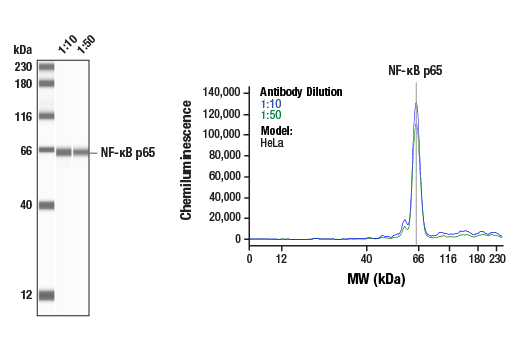
| Simple Western™ | 1:10 - 1:50 |
| Immunoprecipitation | 1:100 |
| Immunohistochemistry (Paraffin) | 1:400 - 1:1600 |
| Immunofluorescence (Immunocytochemistry) | 1:400 - 1:1600 |
| Flow Cytometry (Fixed/Permeabilized) | 1:400 - 1:1600 |
| Chromatin IP | 1:100 |
| Chromatin IP-seq | 1:100 |
| CUT&RUN | 1:100 |
Specificity/Sensitivity
物种反应性:
人, 小鼠, 大鼠, 仓鼠 , 猴, 犬
核转录因子 κB (NF-κB)/Rel 家族的转录因子,在炎症和免疫应答中发挥重要作用 (1,2)。在哺乳动物中有五个家族成员:RelA、c-Rel、RelB、NF-κB1 (p105/p50) 和 NF-κB2 (p100/p52)。p105 和 p100 都可由蛋白酶体经蛋白水解处理后,分别产生 p50 和 p52。Rel 蛋白结合 p50 和 p52 形成二聚体,二聚体再结合 DNA,调节转录活动。未刺激细胞中 NF-κB 被 IκB 抑制性蛋白阻隔在细胞浆中 (3-5)。NF-κB 活化剂可诱导 IκB 蛋白的磷酸化,并通过泛素-蛋白酶体通路将其靶标进行快速降解,释放 NF-κB 进入胞核,使其调节基因表达 (6-8)。NIK 和 IKKα (IKK1) 可调节 NF-κB2 (p100) 的磷酸化和加工以产生 p52,后者再转位入胞核 (9-11)。 Baeuerle, P.A. and Henkel, T. (1994) Annu Rev Immunol 12, 141-79. Baeuerle, P.A. and Baltimore, D. (1996) Cell 87, 13-20. Haskill, S. et al. (1991) Cell 65, 1281-9. Thompson, J.E. et al. (1995) Cell 80, 573-82. Whiteside, S.T. et al. (1997) EMBO J 16, 1413-26. Traenckner, E.B. et al. (1995) EMBO J 14, 2876-83. Scherer, D.C. et al. (1995) Proc Natl Acad Sci USA 92, 11259-63. Chen, Z.J. et al. (1996) Cell 84, 853-62. Senftleben, U. et al. (2001) Science 293, 1495-9. Coope, H.J. et al. (2002) EMBO J 21, 5375-85. Xiao, G. et al. (2001) Mol Cell 7, 401-9. Kozako, T. et al. (2015) Sci Rep 5, 11345.
参考图片
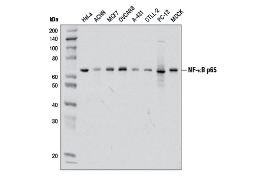
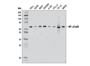
Western blot analysis of extracts from various cell lines using NF-κB p65 (D14E12) XP® Rabbit mAb.Western免疫印迹。用NF-κB p65 (D14E12) XP® Rabbit mAb研究各种类型细胞的细胞提取液。
Confocal immunofluorescent analysis of HT-1080 cells, untreated (left) or treated with hTNF-α #8902 (20 ng/ml, 20 min) (right), using NF-κB p65 (D14E12) XP® Rabbit mAb (green). Actin filaments were labeled with DY-554 phalloidin (red). Blue pseudocolor = DRAQ5® #4084 (fluorescent DNA dye).共聚焦免疫荧光分析HT-1080 细胞核,未经处理 (左图)或 经hTNF-α #8902 (20 ng/ml, 20 min) (右图)处理, 所用抗体为NF-κB p65 (D14E12) XP® Rabbit mAb (绿色) 肌动蛋白微丝用DY-554 phalloidin标记(红色)。Blue pseudocolor=DRAQ5® #4084 DNA 荧光染料。
Chromatin immunoprecipitations were performed with cross-linked chromatin from 4 x 10⁶ HeLa cells treated with hTNF-α #8902 (30 ng/ml, 1 hr) and either 5 μl of NF-κB p65 (D14E12) XP® Rabbit mAb or 2 μl of Normal Rabbit IgG #2729 using SimpleChIP® Enzymatic Chromatin IP Kit (Magnetic Beads) #9003. The enriched DNA was quantified by Real-Time PCR using SimpleChIP® Human IκBα Promoter Primers #5552, human IL-8 promoter primers, and SimpleChIP® Human α Satellite Repeat Primers #4486. The amount of immunoprecipitated DNA in each sample is represented as signal relative to the total amount of input chromatin, which is equivalent to one.染色质免疫共沉淀。HeLa细胞培养至4 x 10⁶,并用hTNF-α #8902 (30 ng/ml, 1 hr)处理后,将染色质交联到玻片上,然后用SimpleChIP® Enzymatic Chromatin IP Kit (Magnetic Beads) #9003进行免疫沉淀实验,本实验中用5 μl of NF-κB p65 (D14E12) XP® Rabbit mAb抗体或2 μl Normal Rabbit IgG #2729 。对富集的DNA做real-time PCR,所用引物为SimpleChIP® Human IκBα Promoter Primers #5552, human IL-8 promoter primers和SimpleChIP® Human α Satellite Repeat Primers #4486。每个样本中沉淀的DNA量定义为相对信号与输入的总染色质相比的数值。
Flow cytometric analysis of HeLa cells using NF-κB p65 (D14E12) XP® Rabbit mAb (blue) compared to concentration matched Rabbit (DA1E) mAb IgG XP® Isotype Control #3900 (red).流式细胞仪研究HeLa细胞,所用抗体为NF-κB p65 (D14E12) XP® Rabbit mAb (蓝色) 浓度相匹配的 Rabbit (DA1E) mAb IgG XP® 同型对照#3900 (红色)。
Immunohistochemical analysis of paraffin-embedded human chronic cholecystitis using NF-κB p65 (D14E12) XP® Rabbit mAb.免疫组织化学染色分析石蜡包埋人慢性胆囊炎组织。所用抗体为NF-κB p65 (D14E12) XP® Rabbit mAb。

 危险品化学品经营许可证(不带存储) 许可证编号:沪(杨)应急管危经许[2022]202944(QY)
  营业执照(三证合一)