


 下载产品说明书
 用小程序,查商品更便捷


 收藏
收藏
 对比
对比 咨询
咨询
Specificity/Sensitivity
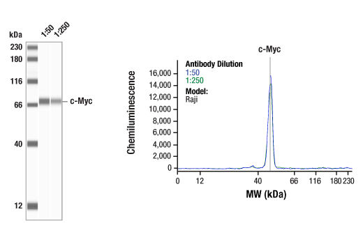
参考图片
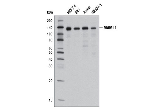
Western blot analysis of extracts from various cell lines using MAML1 (D3K7B) Rabbit mAb.
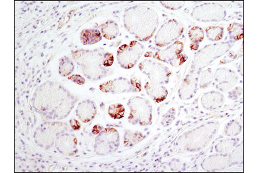
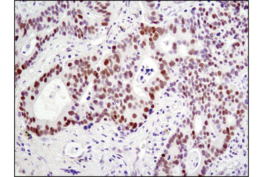
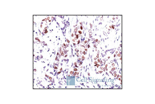
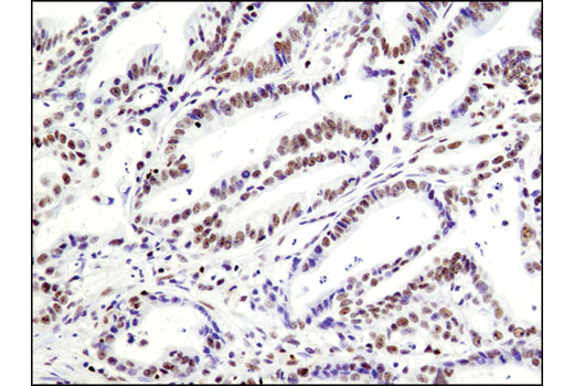
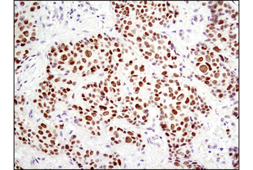
Immunohistochemical analysis of paraffin-embedded human breast carcinoma using HES1 (D6P2U) Rabbit mAb.
Immunohistochemical analysis of paraffin-embedded human lung carcinoma using HES1 (D6P2U) Rabbit mAb.
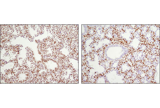
Immunohistochemical analysis of paraffin-embedded CUTLL1 cell pellets, control (left) or Compound E-treated (right), using HES1 (D6P2U) Rabbit mAb.
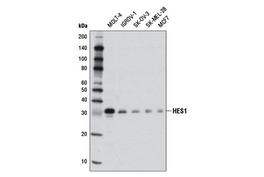
Western blot analysis of extracts from various cell lines using HES1 (D6P2U) Rabbit mAb.
After the primary antibody is bound to the target protein, a complex with HRP-linked secondary antibody is formed. The LumiGLO® is added and emits light during enzyme catalyzed decomposition.
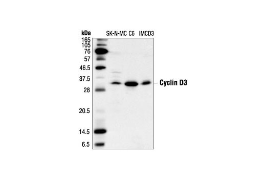
Western blot analysis of extracts from SK-N-MC, C6 and IMCD3 cells, using Cyclin D3 (DCS22) Mouse mAb.
Immunohistochemical analysis of paraffin-embedded human breast carcinoma, using Cyclin D3 (DCS22) Mouse mAb.
Immunohistochemical analysis of paraffin-embedded human lung carcinoma, using Cyclin D3 (DCS22) Mouse mAb.
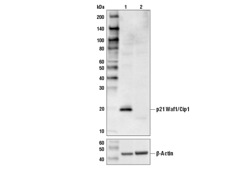
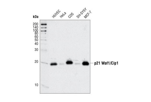
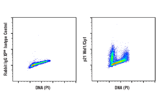
Western blot analysis of extracts from various cell types using p21 Waf1/Cip1 (12D1) Rabbit mAb.
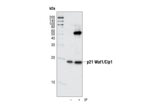
Immunoprecipitation of p21 from human umbillical vein endothelial cells (HUVECs) using p21 Waf1/Cip1 (12D1) Rabbit mAb. Western blot detection was performed using the same antibody.
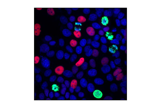
Confocal immunofluorescent analysis of MCF7 cells using p21 Waf1/Cip1 (12D1) Rabbit mAb (red) and Phospho-Histone H3 (Ser10) (6G3) Mouse mAb #9706 (green). Blue pseudocolor = DRAQ5® #4084 (fluorescent DNA dye).
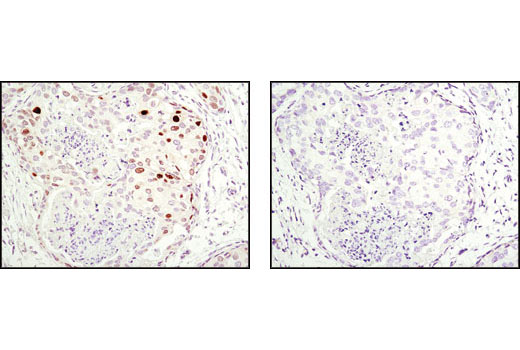
Immunohistochemical analysis of paraffin-embedded human breast carcinoma using p21 Waf1/Cip1 (12D1) Rabbit mAb in the presence of control peptide (left) or antigen-specific peptide (right).
Flow cytometric analysis of HeLa cells (red) and MCF7 cells (blue), using p21 Waf1/Cip1 (12D1) Rabbit mAb.
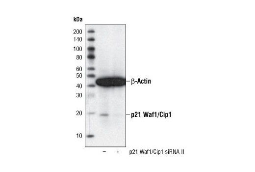
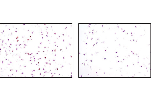
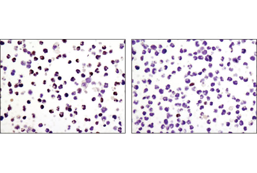
Immunohistochemical analysis of paraffin-embedded HeLa cells, transfected with SignalSilence® Control siRNA (Unconjugated) #6568 (left) or SignalSilence® p21 Waf1/Cip1 siRNA II #6558 (right), using p21 Waf1/Cip1 (12D1) Rabbit mAb.
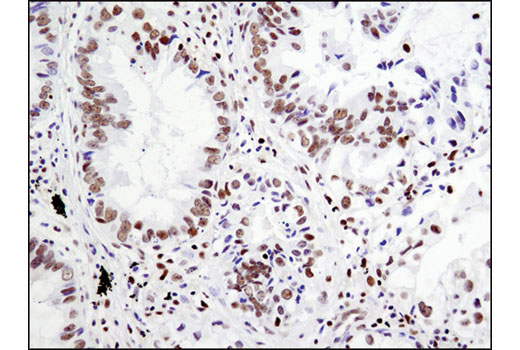
Immunohistochemical analysis of paraffin-embedded human breast carcinoma using Notch1 (D1E11) XP® Rabbit mAb.
Immunohistochemical analysis of paraffin-embedded human inflammatory granulation tissue using Notch1 (D1E11) XP® Rabbit mAb.
Immunohistochemical analysis of paraffin-embedded A2780 (left), Jurkat (center) and RL (right) cell pellets using Notch1 (D1E11) XP® Rabbit mAb. Both A2780 and Jurkat express Notch1, but only Jurkat cells have cleaved Notch1, while RL cells express very low or no Notch1.
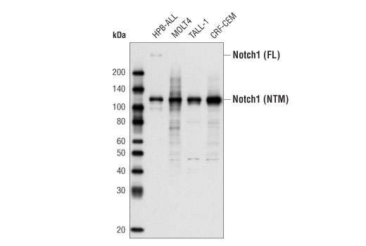
Western blot analysis of total cell extract from various cell types using Notch1 (D1E11) XP® Rabbit mAb. The full-length (FL) Notch protein and the cleaved transmembrane/intracellular region (NTM) are indicated.
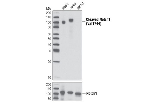
Western blot analysis of extracts from various cell lines using Cleaved Notch1 (Val1744) (D3B8) Rabbit mAb (upper) or Notch1 (D1E11) XP® Rabbit mAb #3608 (lower).
Western blot analysis of extracts from HeLa cells, transfected with 100 nM SignalSilence® Control siRNA (Fluorescein Conjugate) #6201 (-) or SignalSilence® p21 Waf1/Cip1 siRNA II (+), using p21 Waf1/Cip1 (12D1) Rabbit mAb #2947 and α-Tubulin (11H10) Rabbit mAb #2125. The p21 Waf1/Cip1 (12D1) Rabbit mAb confirms silencing of p21 Waf1/Cip1 expression and α-Tubulin (11H10) Rabbit mAb is used to control for loading and specificity of p21 Waf1/Cip1 siRNA.
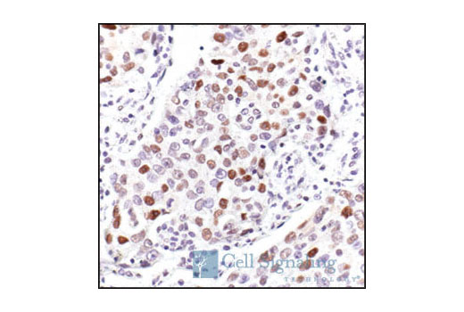
Immunohistochemical analysis of paraffin-embedded human stomach adjacent to MALT (mucosa-associated lymphoid tissue) lymphoma using Notch1 (D1E11) XP® Rabbit mAb.
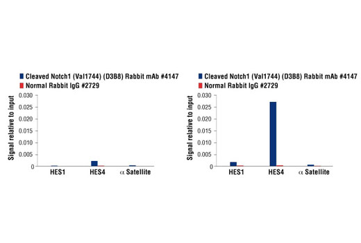
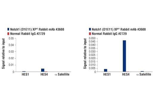
CUTLL1 cells were cultured in media with γ-secretase inhibitor (1μM) for 3 days and then either harvested immediately (left panel) or washed and cultured in fresh media for 3h (right panel). Chromatin immunoprecipitations were performed with cross-linked chromatin from 4 x 10⁶ cells and 2.5 µl of Cleaved Notch1 (Val1744) (D3B8) Rabbit mAb or 2 µl of Normal Rabbit IgG #2729 using SimpleChIP® Enzymatic Chromatin IP Kit (Magnetic Beads) #9003. The enriched DNA was quantified by real-time PCR using human HES1 promoter primers, SimpleChIP® Human HES4 Promoter Primers #7273, and SimpleChIP® Human α Satellite Repeat Primers #4486. The amount of immunoprecipitated DNA in each sample is represented as signal relative to the total amount of input chromatin, which is equivalent to one.
CUTLL1 cells were cultured in media with γ-secretase inhibitor (1 μM) for 3 days and then either harvested immediately (left panel) or washed and cultured in fresh media for 3 hr (right panel). Chromatin immunoprecipitations were performed with cross-linked chromatin from 4 x 10⁶ cells and 5 µl of Notch1 (D1E11) XP® Rabbit mAb or 2 µl of Normal Rabbit IgG #2729 using SimpleChIP® Enzymatic Chromatin IP Kit (Magnetic Beads) #9003. The enriched DNA was quantified by real-time PCR using human HES1 promoter primers, SimpleChIP® Human HES4 Promoter Primers #7273, and SimpleChIP® Human α Satellite Repeat Primers #4486. The amount of immunoprecipitated DNA in each sample is represented as signal relative to the total amount of input chromatin, which is equivalent to one.
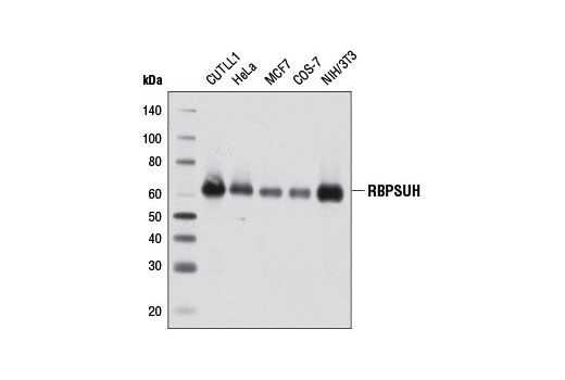
Western blot analysis of extracts from various cell lines using RBPSUH (D10A4) XP® Rabbit mAb.
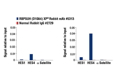
CUTLL1 cells were cultured in media with γ-secretase inhibitor (1 μM, 3 d) and then either harvested immediately (left panel) or washed and cultured in fresh media for 3 h (right panel). Chromatin immunoprecipitations were performed with cross-linked chromatin from 4 x 10⁶ cells and 10 µl of RBPSUH (D10A4) XP® Rabbit mAb or 2 µl of Normal Rabbit IgG #2729 using SimpleChIP® Enzymatic Chromatin IP Kit (Magnetic Beads) #9003. The enriched DNA was quantified by real-time PCR using human HES1 promoter primers, SimpleChIP® Human HES4 Promoter Primers #7273, and SimpleChIP® Human α Satellite Repeat Primers #4486. The amount of immunoprecipitated DNA in each sample is represented as signal relative to the total amount of input chromatin, which is equivalent to one.
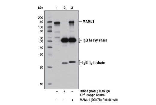
Immunoprecipitation of MAML1 protein from 293T cell extracts, using Rabbit (DA1E) mAb IgG XP® Isotype Control #3900 (lane 2) or MAML1 (D3K7B) Rabbit mAb (lane 3). Lane 1 is 10% input. Western blot analysis was performed using MAML1 Antibody #4608.
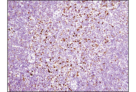
Immunohistochemical analysis of paraffin-embedded mouse lymph node using RBPSUH (D10A4) XP® Rabbit mAb.
Immunohistochemical analysis of paraffin-embedded human lung carcinoma using RBPSUH (D10A4) XP® Rabbit mAb.
Immunohistochemical analysis of paraffin-embedded E18.5 mouse lung, Rbpjk F/+ Shh+/+ (wild type, left) or Rbpjk F/- Shhcre/+ (Rbpjk conditional knock out, right), using RBPSUH (D10A4) XP® Rabbit mAb. Note lack of staining in the bronchial epithelial cells in the conditional knock out tissue (right). Tissue courtesy of Dr. Wellington Cardosa, Boston University School of Medicine.
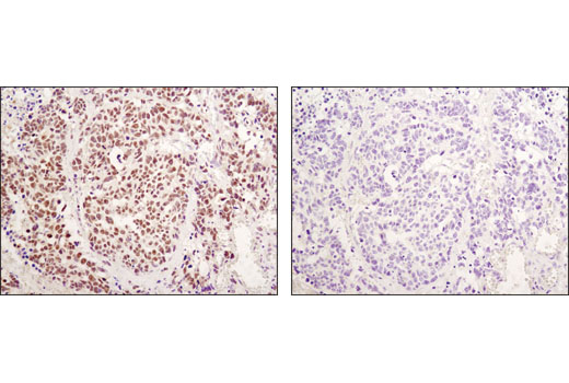
Immunohistochemical analysis of paraffin-embedded human lung carcinoma using RBPSUH (D10A4) XP® Rabbit mAb in the presence of control peptide (left) or antigen-specific peptide (right).
Immunohistochemical analysis of paraffin-embedded human colon carcinoma using RBPSUH (D10A4) XP® Rabbit mAb.
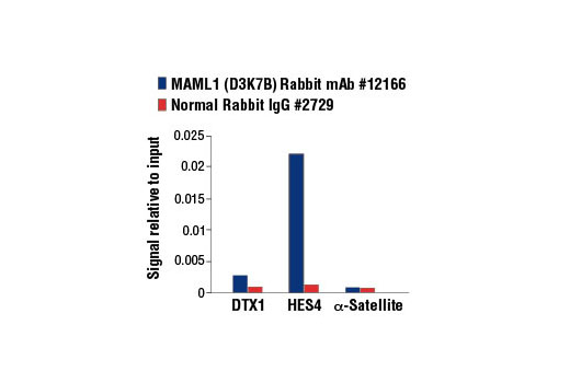
CUTLL1 cells were cultured in media with γ-secretase inhibitor (1 μM) for 3 days and then washed and cultured in fresh media for 3 h. Chromatin immunoprecipitations were performed with cross-linked chromatin from 4 x 10⁶ cells and 5 µl of MAML1 (D3K7B) Rabbit mAb or 2 µl of Normal Rabbit IgG #2729 using SimpleChIP® Enzymatic Chromatin IP Kit (Magnetic Beads) #9003. The enriched DNA was quantified by real-time PCR using human DTX1 intron 3 primers, SimpleChIP® Human HES4 Promoter Primers #7273, and SimpleChIP® Human α Satellite Repeat Primers #4486. The amount of immunoprecipitated DNA in each sample is represented as signal relative to the total amount of input chromatin, which is equivalent to one.
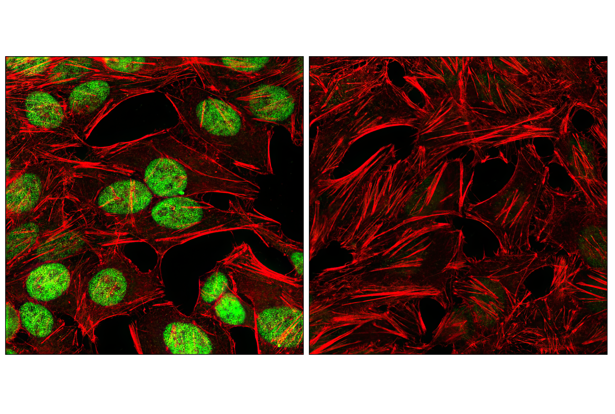
Confocal immunofluorescent analysis of HT-29 cells, untreated (left) or treated with bromodomain inhibitor JQ1 (1 μM, 72 hr; right), using c-Myc (D3N8F) Rabbit mAb (green). Actin filaments were labeled with DyLight™ 554 Phalloidin #13054 (red).
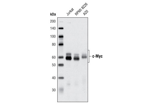
Western blot analysis of extracts from various cell lines using c-Myc (D3N8F) Rabbit mAb.
Western blot analysis of extracts from HT-29 cells, untreated (-) or treated with JQ1 (1 μM; 72 hr; +), using c-Myc (D3N8F) Rabbit mAb (upper) and β-Actin (D6A8) Rabbit mAb #8457 (lower). As expected, the BET bromodomain inhibitor JQ1 inhibits c-Myc expression.
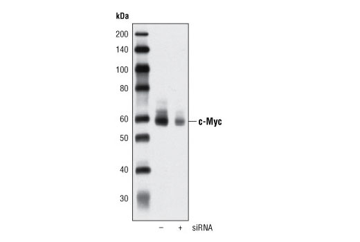
Western blot analysis of extracts from HeLa cells, transfected with 100 nM SignalSilence® Control siRNA (Unconjugated) #6568 (-) or SignalSilence® c-Myc siRNA I #6341 (+), using c-Myc (D3N8F) Rabbit mAb (upper) or β-Actin (D6A8) Rabbit mAb #8457 (lower). The c-Myc (D3N8F) Rabbit mAb confirms silencing of c-Myc expression, while the β-Actin (D6A8) Rabbit mAb is used as a loading control.
Chromatin immunoprecipitations were performed with cross-linked chromatin from 4 x 10⁶ Daudi cells and either 10 μl of c-Myc (D3N8F) Rabbit mAb or 2 μl of Normal Rabbit IgG #2729 using SimpleChIP® Enzymatic Chromatin IP Kit (Magnetic Beads) #9003. The enriched DNA was quantified by real-time PCR using human ATF4 promoter primers, SimpleChIP® Human NPM1 Intron 1 Primers #4779, and SimpleChIP® Human α Satellite Repeat Primers #4486. The amount of immunoprecipitated DNA in each sample is represented as signal relative to the total amount of input chromatin, which is equivalent to one.
Flow cytometric analysis of HT29 cells using Myc Rabbit mAb, treated with JQ1 (1uM, 72 hours at 37C) (blue) and untreated (green). Anti-rabbit IgG (H+L), F(ab')2 Fragment (Alexa Fluor® 488 Conjugate) was used as a secondary antibody.
Western blot analysis of total cell extract from various cell types using Notch1 (D1E11) XP® Rabbit mAb #3608. The full-length (FL) Notch protein and the cleaved transmembrane/intracellular region (NTM) are indicated.
Western blot analysis of extracts from various cell lines using Cleaved Notch1 (Val1744) (D3B8) Rabbit mAb #4147 (upper) or Notch1 (D1E11) XP® Rabbit mAb #3608 (lower).
Western blot analysis of extracts from various cell lines using RBPSUH (D10A4) XP® Rabbit mAb #5313. (D10A4) XP® Rabbit mAb #5313.
Western blot analysis of extracts from various cell lines using MAML1 (D3K7B) Rabbit mAb #12166.
Western blot analysis of extracts from various cell lines using c-Myc (D3N8F) Rabbit mAb #13987.
Western blot analysis of extracts from various cell types using p21 Waf1/Cip1 (12D1) Rabbit mAb #2947.
Western blot analysis of extracts from various cell lines using HES1 (D6P2U) Rabbit mAb #11988.
Western blot analysis of extracts from SK-N-MC, C6 and IMCD3 cells, using Cyclin D3 (DCS22) Mouse mAb #2936.
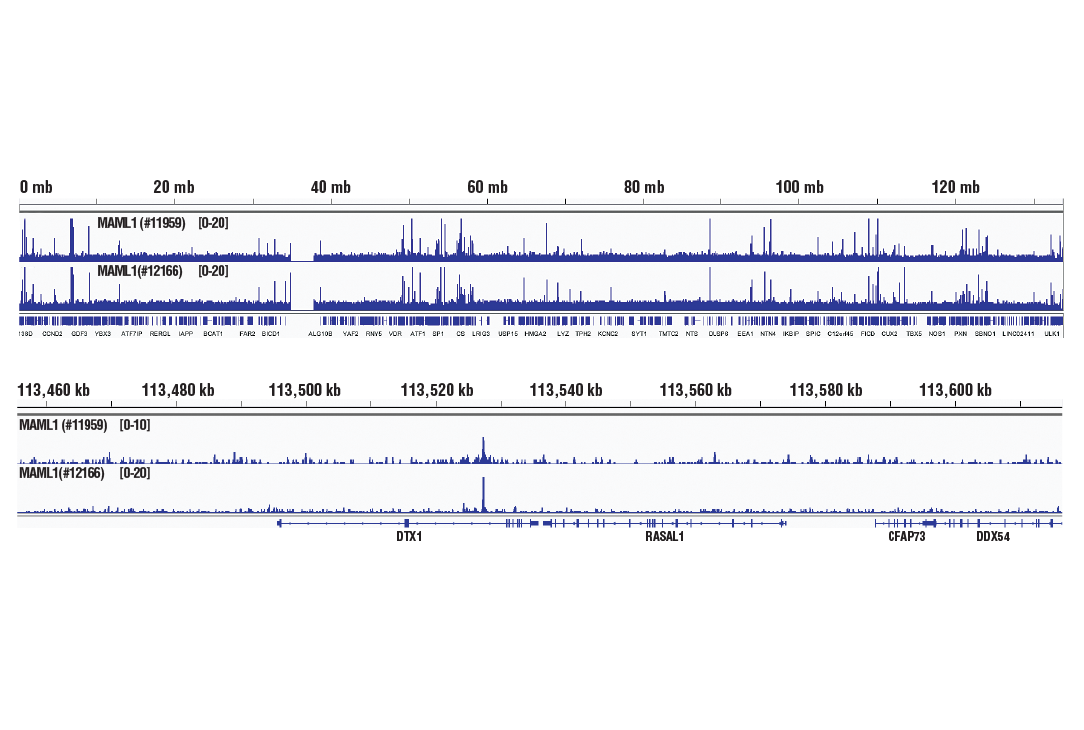
Chromatin immunoprecipitations were performed with cross-linked chromatin from 4 x 10⁶ Daudi cells and 10 μl of c-Myc (D3N8F) Rabbit mAb, using SimpleChIP® Enzymatic Chromatin IP Kit (Magnetic Beads) #9003. DNA Libraries were prepared from 5 ng enriched ChIP DNA using NEBNext® Ultra™ II DNA Library Prep Kit for Illumina®, and sequenced on the Illumina NextSeq. The figure shows binding across NPM1, a known target gene of c-Myc (see additional figure containing ChIP-qPCR data). For additional ChIP-seq tracks, please download the product data sheet.

 危险品化学品经营许可证(不带存储) 许可证编号:沪(杨)应急管危经许[2022]202944(QY)
  营业执照(三证合一)