


 下载产品说明书
 用小程序,查商品更便捷


 收藏
收藏
 对比
对比 咨询
咨询
Specificity/Sensitivity
参考图片
Western blot analysis of extracts from various cell lines using Jagged2 (C23D2) Rabbit mAb #2210.
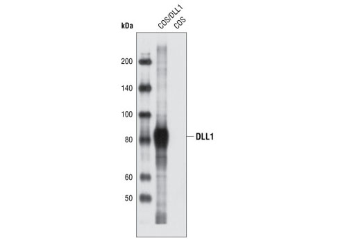
Western blot analysis of extracts from COS cells, untransfected or transiently transfected with a construct expressing rat DLL1 protein, using DLL1 Antibody #2588.
After the primary antibody is bound to the target protein, a complex with HRP-linked secondary antibody is formed. The LumiGLO* is added and emits light during enzyme catalyzed decomposition.
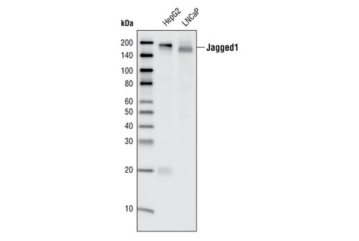
Western blot analysis of total cell lysates from HepG2 and LNCaP cells, using Jagged1 (28H8) Rabbit mAb.
Western blot analysis of total cell lysates from HeLa, SK-OV-3 and SR cells using Jagged2 (C23D2) Rabbit mAb.
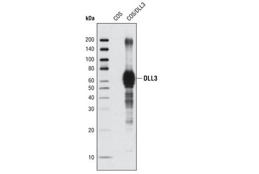
Western blot analysis of COS cell extracts, untransfected or transiently transfected with a construct expressing rat DLL3 protein, using DLL3 (G93) Antibody.
Western blot analysis of extracts from COS cells, untransfected or transiently transfected with a construct expressing rat DLL1 protein, using DLL1 Antibody.
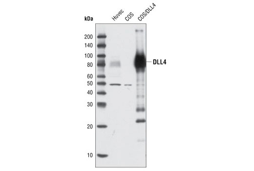
Western blot analysis of extracts from HUVEC and COS cells, untransfected or transiently transfected with a construct expressing human DLL4, using DLL4 Antibody.
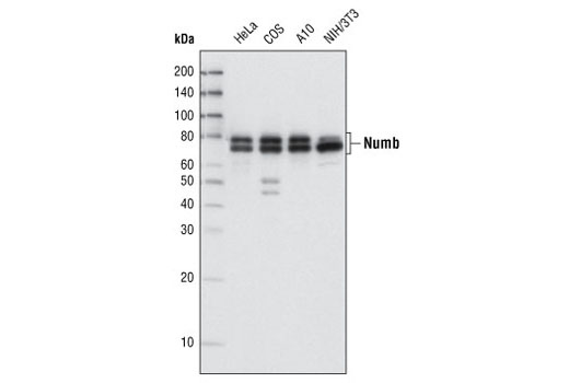
Western blot analysis of extracts from various cell lines using Numb (C29G11) Rabbit mAb.
Flow cytometric analysis of A-204 cells using Numb (C29G11) Rabbit mAb (blue) compared to a nonspecific negative control antibody (red).
Confocal immunofluorescent analysis of HeLa cells using Numb (C29G11) Rabbit mAb (green). Actin filaments have been labeled with DY-554 phalloidin (red). Blue pseudocolor = DRAQ5® #4084 (fluorescent DNA dye).
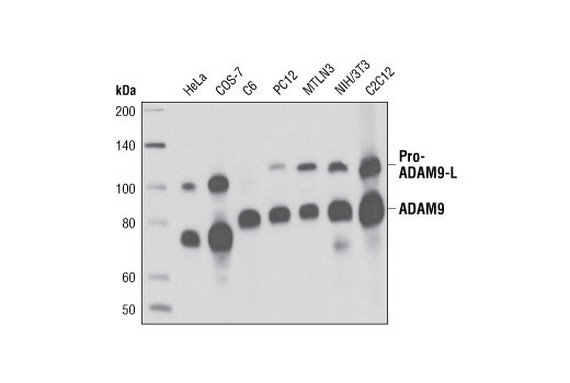
Western blot analysis of extracts from various cell types using ADAM9 (D64B5) Rabbit mAb.
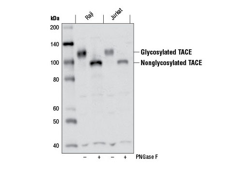
Western blot analysis of extracts from Raji and Jurkat cells, untreated or treated with peptide N-glycosidase F (PNGase F), using TACE (D22H4) Rabbit mAb.
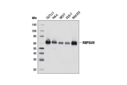
Western blot analysis of extracts from various cell lines using RBPSUH (D10A4) XP® Rabbit mAb.
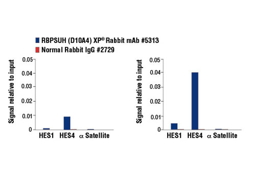
CUTLL1 cells were cultured in media with γ-secretase inhibitor (1 μM, 3 d) and then either harvested immediately (left panel) or washed and cultured in fresh media for 3 h (right panel). Chromatin immunoprecipitations were performed with cross-linked chromatin from 4 x 10⁶ cells and 10 µl of RBPSUH (D10A4) XP® Rabbit mAb or 2 µl of Normal Rabbit IgG #2729 using SimpleChIP® Enzymatic Chromatin IP Kit (Magnetic Beads) #9003. The enriched DNA was quantified by real-time PCR using human HES1 promoter primers, SimpleChIP® Human HES4 Promoter Primers #7273, and SimpleChIP® Human α Satellite Repeat Primers #4486. The amount of immunoprecipitated DNA in each sample is represented as signal relative to the total amount of input chromatin, which is equivalent to one.
Western blot analysis of extracts from HepG2 and LNCaP cells using Jagged1 (28H8) Rabbit mAb #2620.
Western blot analysis of extracts from COS cells, untransfected or transiently transfected with a construct expressing rat DLL3 protein, using DLL3 (G93) Antibody #2483.
Western blot analysis of extracts from HUVEC and COS cells, untransfected or transiently transfected with a construct expressing human DLL4, using DLL4 Antibody #2589.
Western blot analysis of extracts from various cell lines using ADAM9 (D64B5) Rabbit mAb #4151.
Western blot analysis of extracts from Raji and Jurkat cells, untreated (-) or treated with peptide N-glycosidase F (PNGase F; +), using TACE (D22H4) Rabbit mAb #6978.
Western blot analysis of extracts from various cell lines using Numb (C29G11) Rabbit mAb #2756.
Western blot analysis of extracts from various cell lines using RBPSUH (D10A4) XP® Rabbit mAb #5313.
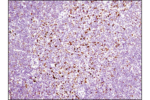
Immunohistochemical analysis of paraffin-embedded mouse lymph node using RBPSUH (D10A4) XP® Rabbit mAb.
Immunohistochemical analysis of paraffin-embedded human lung carcinoma using RBPSUH (D10A4) XP® Rabbit mAb.
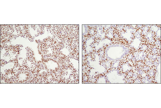
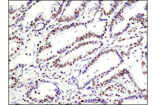
Immunohistochemical analysis of paraffin-embedded E18.5 mouse lung, Rbpjk F/+ Shh+/+ (wild type, left) or Rbpjk F/- Shhcre/+ (Rbpjk conditional knock out, right), using RBPSUH (D10A4) XP® Rabbit mAb. Note lack of staining in the bronchial epithelial cells in the conditional knock out tissue (right). Tissue courtesy of Dr. Wellington Cardosa, Boston University School of Medicine.
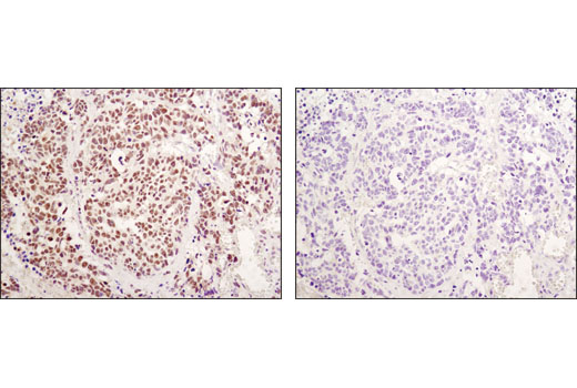
Immunohistochemical analysis of paraffin-embedded human lung carcinoma using RBPSUH (D10A4) XP® Rabbit mAb in the presence of control peptide (left) or antigen-specific peptide (right).
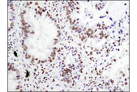
Immunohistochemical analysis of paraffin-embedded human colon carcinoma using RBPSUH (D10A4) XP® Rabbit mAb.

 危险品化学品经营许可证(不带存储) 许可证编号:沪(杨)应急管危经许[2022]202944(QY)
  营业执照(三证合一)