


 用小程序,查商品更便捷


WB
1:500IHC-P
1:1000FC(Intra)
1:250IP
1:25
PARP1 possesses a nuclear localization sequence (NLS) near the DNA-binding domain and a caspase-cleavage site between the DNA-binding domain and the automodification domain [PMID: 10455009]. During caspasedependent apoptosis, PARP1 is cleaved by caspases 3 and 7 at its caspase-cleavage site into 24-kDa and 89-kDa fragments [PMID: 10497198, PMID: 8358726]. The 24-kDa PARP1 fragment contains the DNAbinding motif and the NLS, whereas the 89-kDa PARP1 fragment contains the automodification and catalytic domains. After PARP1 cleavage by caspase, the 24-kDa PARP1 fragment irreversibly binds to DNA breaks and acts as a transdominant inhibitor of active PARP1, whereas the 89-kDa PARP1 fragment is translocated to the cytoplasm [PMID: 11570811, PMID: 9721847]. Thereby, PARP1 fragmentation by caspase leads to its inactivation, which inhibits DNA repair and facilitates caspase-mediated DNA fragmentation in apoptosis.
12 months from date of receipt, 4°C as supplied
参考图片
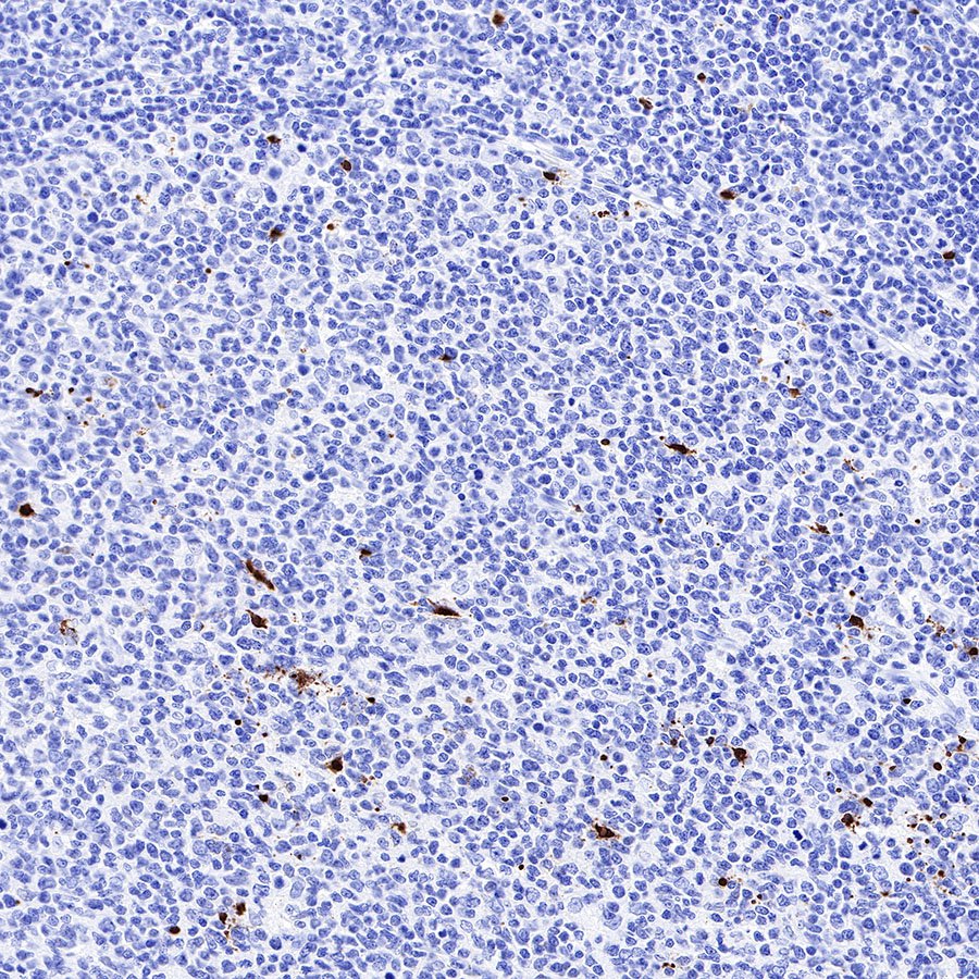
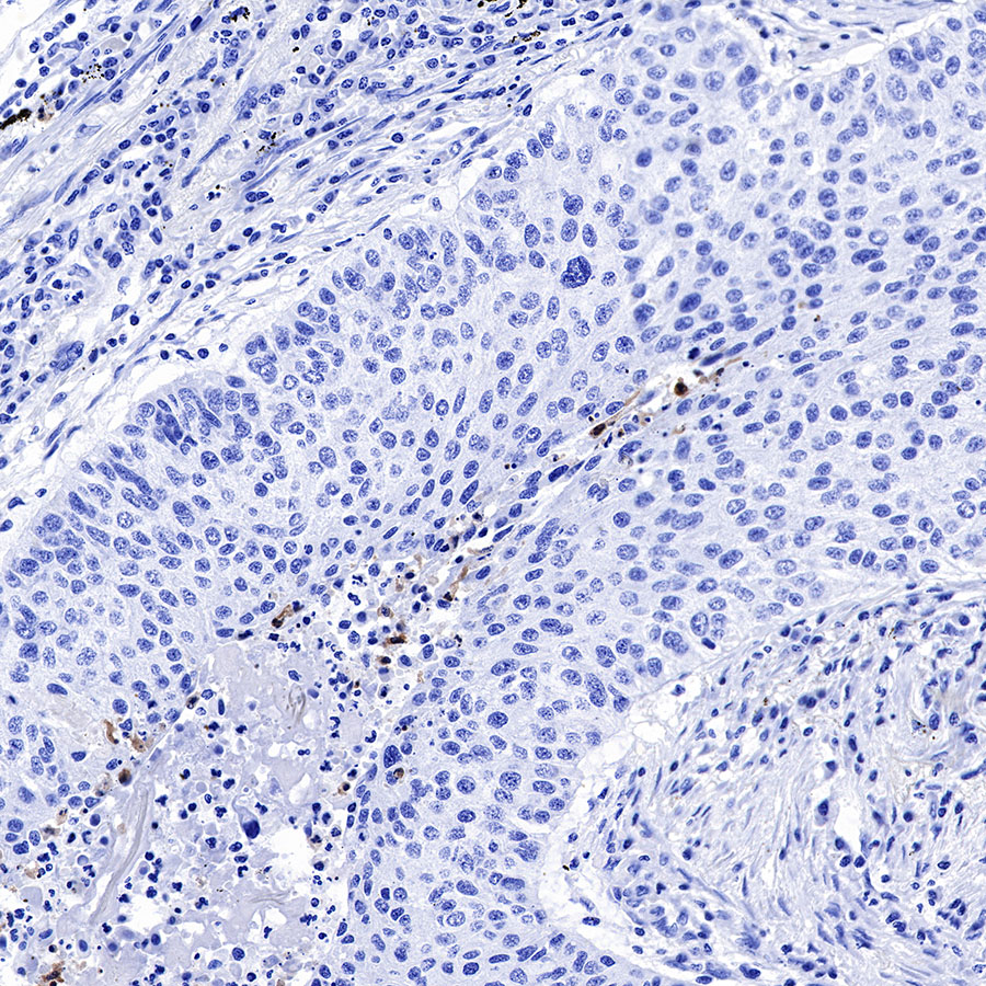
IHC shows positive staining in paraffin-embedded human tonsil. Anti-Cleaved PARP1 antibody was used at 1/1000 dilution, followed by a HRP Polymer for Mouse & Rabbit IgG (ready to use). Counterstained with hematoxylin. Heat mediated antigen retrieval with Tris/EDTA buffer pH9.0 was performed before commencing with IHC staining protocol.
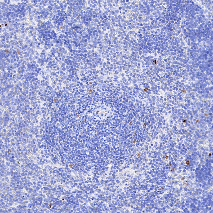
IHC shows positive staining in paraffin-embedded mouse spleen. Anti-Cleaved PARP1 antibody was used at 1/1000 dilution, followed by a HRP Polymer for Mouse & Rabbit IgG (ready to use). Counterstained with hematoxylin. Heat mediated antigen retrieval with Tris/EDTA buffer pH9.0 was performed before commencing with IHC staining protocol.
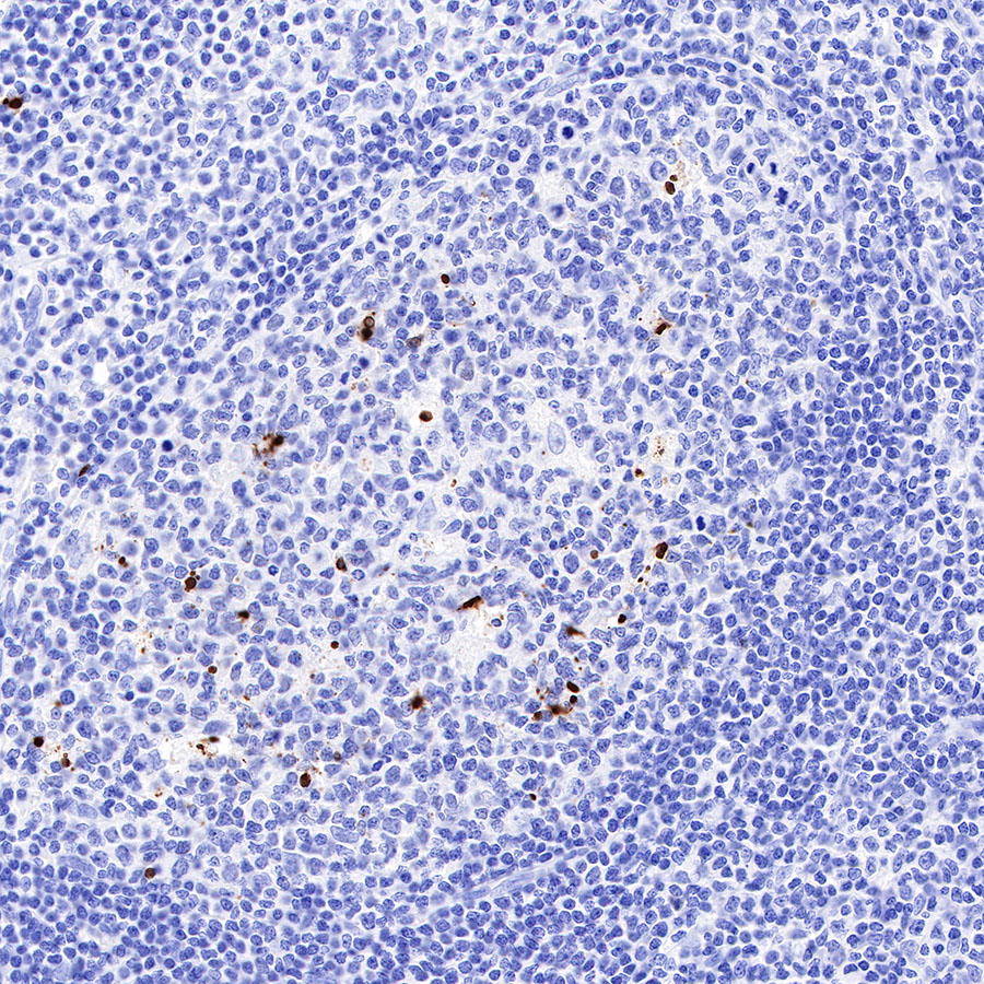
IHC shows positive staining in paraffin-embedded human tonsil. Anti-Cleaved PARP1 antibody was used at 1/1000 dilution, followed by a HRP Polymer for Mouse & Rabbit IgG (ready to use). Counterstained with hematoxylin. Heat mediated antigen retrieval with Tris/EDTA buffer pH9.0 was performed before commencing with IHC staining protocol.
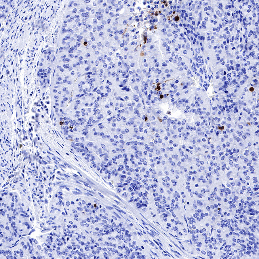
IHC shows positive staining in paraffin-embedded human endometrial cancer. Anti-Cleaved PARP1 antibody was used at 1/1000 dilution, followed by a HRP Polymer for Mouse & Rabbit IgG (ready to use). Counterstained with hematoxylin. Heat mediated antigen retrieval with Tris/EDTA buffer pH9.0 was performed before commencing with IHC staining protocol.
IHC shows positive staining in paraffin-embedded human lung cancer. Anti-Cleaved PARP1 antibody was used at 1/1000 dilution, followed by a HRP Polymer for Mouse & Rabbit IgG (ready to use). Counterstained with hematoxylin. Heat mediated antigen retrieval with Tris/EDTA buffer pH9.0 was performed before commencing with IHC staining protocol.
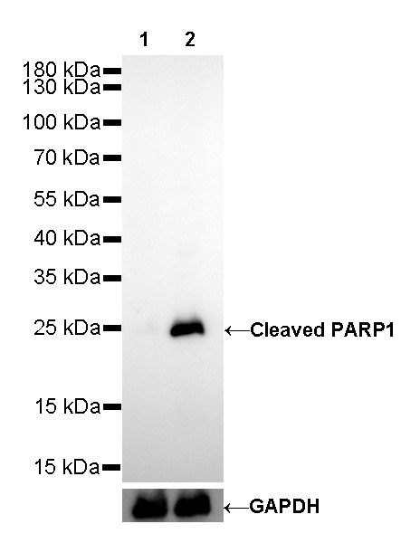
WB result of Cleaved PARP1 Rabbit mAb
Primary antibody: Cleaved PARP1 Rabbit mAb at 1/500 dilution
Lane 1: Untreated HeLa whole cell lysate 20 µg
Lane 2: HeLa treated with Staurosporine (1 μM, 3 hr) whole cell lysate 20 µg
Negative control: Untreated HeLa whole cell lysate
Secondary antibody: Goat Anti-Rabbit IgG, (H+L), HRP conjugated at 1/10000 dilution
Predicted MW: 24 kDa
Observed MW: 24 kDa
Exposure time: 30s
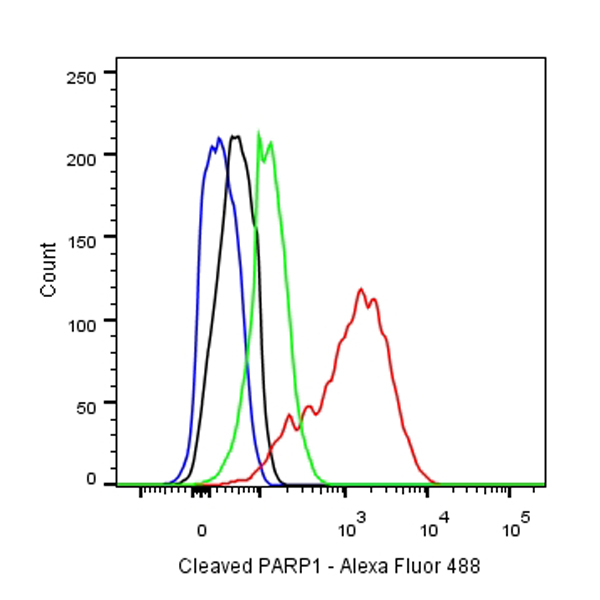
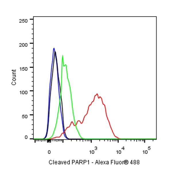
Flow cytometric analysis of HeLa treated with 1 μM Staurosporine for 3 hours labelling Cleaved PARP1 antibody at 1/250 (0.1 μg) dilution/(red)/Untreated control cells labelling Cleaved PARP1 antibody at 1/250 (0.1 μg)/(Green) compared with a Rabbit monoclonal IgG (Black) isotype control and an unlabelled control (cells without incubation with primary antibody and secondary antibody) (Blue). Goat Anti-Rabbit IgG Alexa Fluor® 488 was used as the secondary antibody.
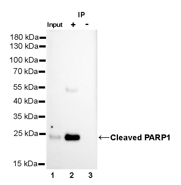
Cleaved PARP1 Rabbit mAb at 1/25 dilution (1µg) immunoprecipitating Cleaved PARP1 in 0.4mg HeLa treated with Staurosporine (1 μM, 3 hr) whole cell lysate.
Western blot was performed on the immunoprecipitate using Cleaved PARP1 Rabbit mAb at 1/1000 dilution.
Secondary antibody (HRP) for IP was used at 1/400 dilution.
Lane 1 : HeLa treated with Staurosporine (1 μM, 3 hr) whole cell lysate 10µg(input)
Lane 2 : Cleaved PARP1 Rabbit mAb IP in HeLa treated with Staurosporine (1 μM, 3 hr) whole cell lysate
Lane 3 : Rabbit monoclonal IgG IP in HeLa treated with Staurosporine (1 μM, 3 hr) whole cell lysate
Predicted MW: 24 kDa
Observed MW: 24 kDa
Exposure time: 60s
Intracellular flow cytometric analysis of 4% PFA fixed 90% methanol permeabilized HeLa (Human cervix adenocarcinoma epithelial cell) cells, treated with 1 μM Staurosporine for 3h (Red) or untreated (Green), labeling Cleaved PARP1 at 1/250 dilution (0.1 μg) compared with a rabbit monoclonal IgG isotype control (black) and an unlabeled control (cells without incubation with primary antibody and secondary antibody) (Blue). Goat Anti - Rabbit IgG Alexa Fluor® 488 was used as the secondary antibody.

 危险品化学品经营许可证(不带存储) 许可证编号:沪(杨)应急管危经许[2022]202944(QY)
  营业执照(三证合一)