


 下载产品说明书 下载SDS
 用小程序,查商品更便捷


 收藏
收藏
 对比
对比 咨询
咨询Flow Cytometry(0.25 µg/106 cells)
Immunohistochemistry(5-15 µg/mL)
Flow Cytometry(0.25 µg/106 cells)
Immunohistochemistry(5-15 µg/mL)

Leu25-Gln167
Accession # Q02242
Scientific Data
 View Larger
Detection of Mouse PD‑1 by Western Blot. Western blot shows lysates of EL-4 mouse lymphoblast cell line and CTLL-2 mouse cytotoxic T cell line (negative control). PVDF membrane was probed with 0.2 µg/mL of Goat Anti-Mouse PD-1 Antigen Affinity-purified Polyclonal Antibody (Catalog # AF1021) followed by HRP-conjugated Anti-Goat IgG Secondary Antibody (Catalog # HAF017). A specific band was detected for PD-1 at approximately 55 kDa (as indicated). This experiment was conducted under reducing conditions and using Immunoblot Buffer Group 1.
 View Larger
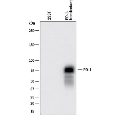
Detection of Mouse PD‑1 by Western Blot. Western blot shows lysates of 293T human embryonic kidney cell line mock transfected or transfected with mouse PD-1. PVDF membrane was probed with 0.2 µg/mL of Goat Anti-Mouse PD-1 Antigen Affinity-purified Polyclonal Antibody (Catalog # AF1021) followed by HRP-conjugated Anti-Goat IgG Secondary Antibody (Catalog # HAF017). A specific band was detected for PD-1 at approximately 75 kDa (as indicated). This experiment was conducted under reducing conditions and using Immunoblot Buffer Group 1.
.") View Larger
View LargerPD‑1 in Mouse Thymus. PD‑1 was detected in perfusion fixed frozen sections of mouse thymus using 15 µg/mL Mouse PD‑1 Antigen Affinity-purified Polyclonal Antibody (Catalog # AF1021) overnight at 4 °C. Tissue was stained (red) and counterstained (green). View our protocol for Fluorescent IHC Staining of Frozen Tissue Sections.
.") View Larger
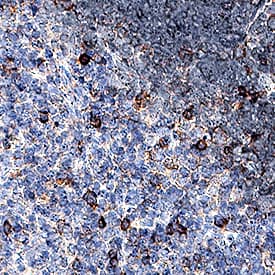
View LargerPD‑1 in Mouse Spleen. PD-1 was detected in perfusion fixed frozen sections of mouse spleen using Goat Anti-Mouse PD-1 Antigen Affinity-purified Polyclonal Antibody (Catalog # AF1021) at 5 µg/mL overnight at 4 °C. Tissue was stained using the Anti-Goat HRP-DAB Cell & Tissue Staining Kit (brown; Catalog # CTS008) and counterstained with hematoxylin (blue). Specific staining was localized to cytoplasm in splenocytes. View our protocol for Chromogenic IHC Staining of Frozen Tissue Sections.
.") View Larger
View LargerPD‑1 in Mouse Thymus. PD-1 was detected in perfusion fixed paraffin-embedded sections of mouse thymus using Goat Anti-Mouse PD-1 Antigen Affinity-purified Polyclonal Antibody (Catalog # AF1021) at 1.7 µg/mL for 1 hour at room temperature followed by incubation with the Anti-Goat IgG VisUCyte™ HRP Polymer Antibody (Catalog # VC004). Before incubation with the primary antibody, tissue was subjected to heat-induced epitope retrieval using Antigen Retrieval Reagent-Basic (Catalog # CTS013). Tissue was stained using DAB (brown) and counterstained with hematoxylin (blue). Specific staining was localized to cell membranes. View our protocol for IHC Staining with VisUCyte HRP Polymer Detection Reagents.
Mouse PD-1 Antibody Summary
Leu25-Gln167
Accession # Q02242
Applications
Please Note: Optimal dilutions should be determined by each laboratory for each application. General Protocols are available in the Technical Information section on our website.
Flow Cytometry(0.25 µg/106 cells)
Immunohistochemistry(5-15 µg/mL)
Background: PD-1
Programmed Death-1 (PD-1) is type I transmembrane protein belonging to the CD28/CTLA-4 family of immunoreceptors that mediate signals for regulating immune responses (1). Other members of this family include CD28, CTLA-4, and ICOS (2-4). PD-1 is most closely related to CTLA-4 and shares approximately 24% amino acid (aa) sequence identity. The mouse PD-1 gene encodes a 288 aa protein with a putative 20 aa signal peptide, a 149 aa extracellular region with one immunoglobulin-like V-type domain, a 21 aa transmembrane domain, and a 98 aa cytoplasmic region. The cytoplasmic tail contains two tyrosine residues that form the immunoreceptor tyrosine-based inhibitory motif (ITIM) and immunoreceptor tyrosine-based switch motif (ITSM) that are important in mediating PD-1 signaling. Mouse and human PD-1 share approximately 69% aa sequence identity. Two B7 family proteins, PD-L1 (also called B7-H1) and PD-L2, have been identified as PD-1 ligands (5, 6). PD-1 is expressed on activated T cells, B cells, myeloid cells, and on a subset of thymocytes. PD-1 deficient mice have a defect in peripheral tolerance and spontaneously develop autoimmune diseases. Binding of PD-1 to PD-L1 or PD-L2 results in the inhibition of TCR-mediated proliferation and cytokine production as well as BCR-mediated signaling. PD-1 likely has an inhibitory role in regulating immune responses (1-4).
- Ishida, Y. et al. (1992) EMBO J. 11:3887.
- Sharpe, A.H. and G.J. Freeman (2002) Nat. Rev. Immunol. 2:116.
- Coyle, A. and J. Gutierrez-Ramos (2001) Nat. Immunol. 2:203.
- Nishimura, H. and T. Honjo (2001) Trends in Immunol. 22:265.
- Latchman Y. et al. (2001) Nature Immun. 2:261.
- Tamura, H. et al. (2001) Blood 97:1809.
Preparation and Storage
- 12 months from date of receipt, -20 to -70 °C as supplied.
- 1 month, 2 to 8 °C under sterile conditions after reconstitution.
- 6 months, -20 to -70 °C under sterile conditions after reconstitution.
参考图片
PD‑1 in Mouse Thymus. PD‑1 was detected in perfusion fixed frozen sections of mouse thymus using 15 µg/mL Mouse PD‑1 Antigen Affinity-purified Polyclonal Antibody (Catalog # AF1021) overnight at 4 °C. Tissue was stained (red) and counterstained (green). View our protocol for Fluorescent IHC Staining of Frozen Tissue Sections.
PD‑1 in Mouse Spleen. PD‑1 was detected in perfusion fixed frozen sections of mouse spleen using Goat Anti-Mouse PD‑1 Antigen Affinity-purified Polyclonal Antibody (Catalog # AF1021) at 5 µg/mL overnight at 4 °C. Tissue was stained using the Anti-Goat HRP-DAB Cell & Tissue Staining Kit (brown; Catalog # CTS008) and counterstained with hematoxylin (blue). Specific staining was localized to cytoplasm in splenocytes. View our protocol for Chromogenic IHC Staining of Frozen Tissue Sections.

 危险品化学品经营许可证(不带存储) 许可证编号:沪(杨)应急管危经许[2022]202944(QY)
  营业执照(三证合一)