


 下载产品说明书 下载SDS
 用小程序,查商品更便捷


 收藏
收藏
 对比
对比 咨询
咨询Product Summary
Precision
Cell Culture Supernates, Cell Lysates, Serum, EDTA Plasma, Heparin Plasma, Urine
| Intra-Assay Precision | Inter-Assay Precision | |||||
|---|---|---|---|---|---|---|
| Sample | 1 | 2 | 3 | 1 | 2 | 3 |
| n | 20 | 20 | 20 | 20 | 20 | 20 |
| Mean (pg/mL) | 125 | 319 | 655 | 131 | 304 | 626 |
| Standard Deviation | 3.49 | 5.6 | 10.8 | 7.36 | 14.6 | 25.8 |
| CV% | 2.6 | 1.8 | 1.6 | 5.6 | 4.8 | 4.1 |
Recovery
The recovery of human PD-1 spiked to levels throughout the range of the assay was evaluated.
| Sample Type | Average % Recovery | Range % |
|---|---|---|
| Cell Culture Media (n=4) | 100 | 97-103 |
| Cell Lysis Buffer (n=1) | 96 | 94-97 |
| EDTA Plasma (n=4) | 86 | 81-93 |
| Heparin Plasma (n=4) | 85 | 75-93 |
| Serum (n=4) | 88 | 77-98 |
| Urine (n=4) | 89 | 83-97 |
Linearity

Scientific Data

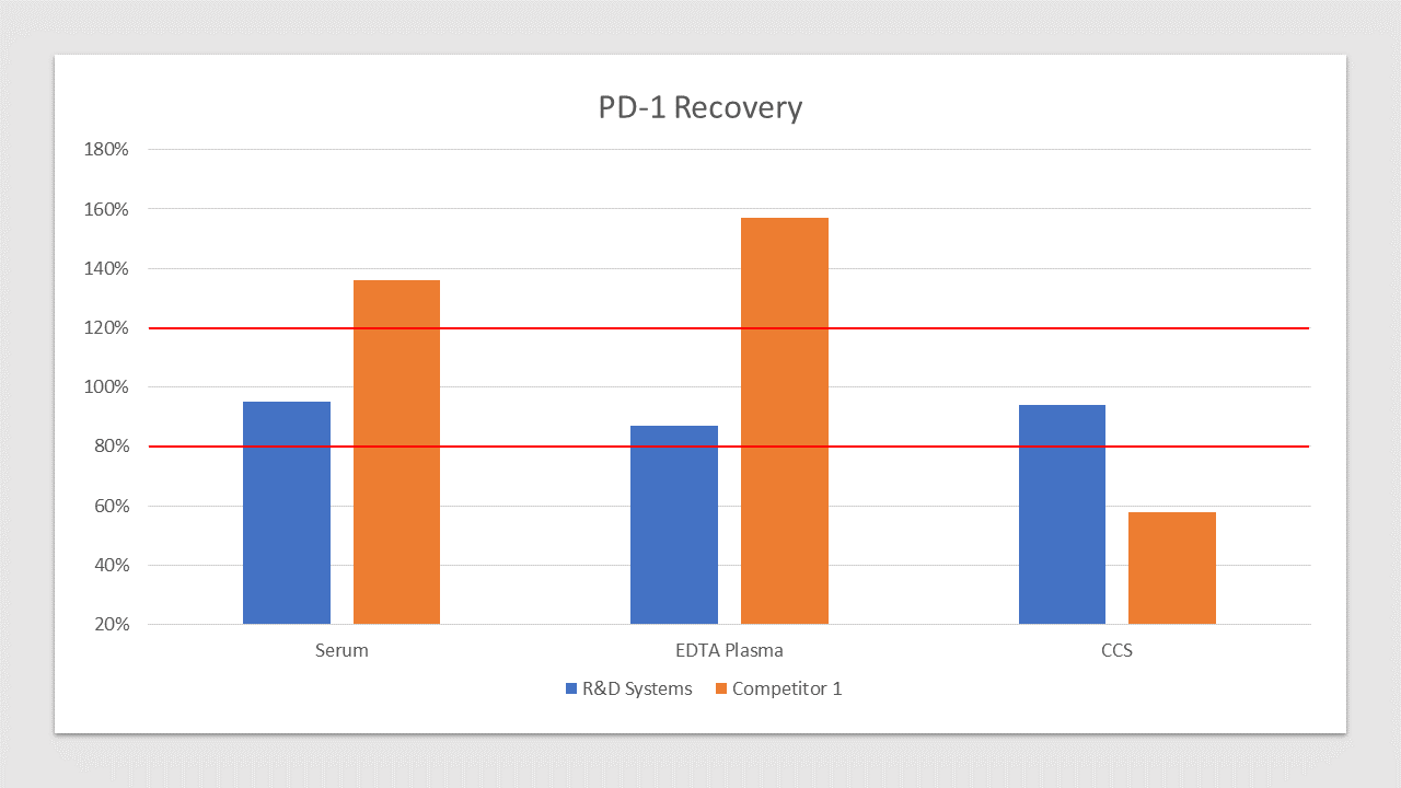
 View Larger
Human PD-1 Quantikine Recovery Competitor Comparison PD-1 is spiked at three known concentrations throughout the range of the assay and run to measure response of the spiked sample matrix. Serum recovery is 95% compared to 136% for the top competitor. EDTA Plasma recovery is 87% compared to 157% for the top competitor. Culture media recovery is 94% compared to 58% for the top competitor. In spike and recovery experiments, natural samples are spiked with the recombinant target analyte of interest to identify interference caused by sample matrices.
Assay Procedure
Refer to the product datasheet for the complete assay procedure.
Bring all reagents and samples to room temperature before use. It is recommended that all samples, standards, and controls be assayed in duplicate.
- Prepare all reagents, standard dilutions, and samples as directed in the product insert.
- Remove excess microplate strips from the plate frame, return them to the foil pouch containing the desiccant pack, and reseal.
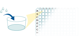
- Add 50 µL of Assay Diluent to each well.
50 µL Assay Diluent
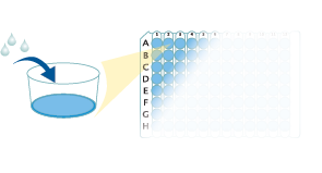
- Add 50 µL of Standard, control, or sample to each well. Cover with a plate sealer, and incubate at room temperature for 2 hours on a horizontal orbital microplate shaker.
- Aspirate each well and wash, repeating the process 3 times for a total of 4 washes.
50 µL Standard, Control, or Sample
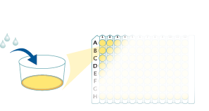
- Add 200 µL of Human PD-1 Conjugate to each well. Cover with a new plate sealer, and incubate at room temperature for 2 hours.
- Aspirate and wash 4 times.
200 µL Conjugate
- Add 200 µL Substrate Solution to each well. Incubate for 30 minutes on the benchtop. Protect from light.
200 µL Substrate Solution
- Add 50 µL of Stop Solution to each well. Read at 450 nm within 30 minutes. Set wavelength correction to 540 nm or 570 nm.
50 µL Stop Solution
Human PD-1 Quantikine ELISA Kit Summary

Background: PD-1
Programmed Death-1 (PD-1), also known as Programmed cell death protein and CD279, is an extensively studied immune checkpoint inhibitory receptor. Given PD-1's role in peripheral tolerance, it is not surprising that increased PD-1 expression is a mechanism for immune escape, which is permissive for cancer growth and metastasis (1,2). PD‐1 is encoded by the PDCD1 gene (3). The PD-1 glycoprotein is a monomeric 50-55kDa type 1 transmembrane protein that belongs to the immunoglobulin (Ig) superfamily (4). PD-1 expression and induction have been well studied (3,5,6). It is expressed in CD4+ and CD8+ T cells as well as B cells, macrophages, some dendritic cell subsets and NK cells. PD-1 is induced by T cell receptor (TCR) signaling as well as interleukin 2 (IL-2), IL-7 and type 1 interferons. A variety of reagents are used to experimentally induce PD-1 expression, including, phorbol 12-myristate 13 acetate, ionomycin, concanavalin A, CD3/CD28 antibodies, or most notably lymphocytic choriomeningitis virus in vivo (6).

 危险品化学品经营许可证(不带存储) 许可证编号:沪(杨)应急管危经许[2022]202944(QY)
  营业执照(三证合一)