


首页
PHENOVUE FLUOR 594 - WGA


品牌: Revvity
 下载SDS
下载SDS 用小程序,查商品更便捷


 收藏
收藏
 对比
对比 咨询
咨询
产品介绍
产品信息
Overview
WGA is a plant lectin that binds different carbohydrate motifs and displays high affinity for sialic acid and N-acetylglucosamine residues of glycoproteins and glycolipids present at the cellular plasma membranes.
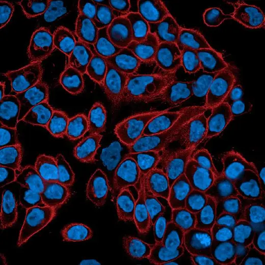
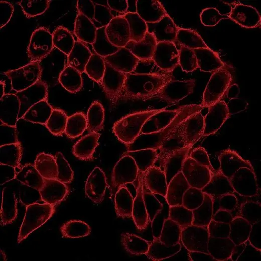
Fluorescent WGA derivatives are commonly used for staining the cellular membranes of mammalian cells, particularly Golgi apparatus which is glycoprotein-enriched. In addition, fluorescent WGA derivatives are also routinely used for the staining of skeletal muscle cells and tissues, as well as in cardiac fibrosis.
PhenoVue Fluor 594 - WGA can be used to visualize cellular membranes in immunofluorescence, immunohistochemistry and flow cytometry, as well as high-content analysis and screening applications.
Additional product information
Features
| Numbers of vials per unit | 5 |
|---|---|
| Quantity or Volume Per Vial | 1mg (29.2nmoles) |
| Form | Lyophilized |
| Storage | 2-8°C |
| Recommended working concentration | 5 µg/mL (146nM) |
| Maximum excitation wavelength | 590 nm |
| Maximum emission wavelength | 617 nm |
| Common filter set | Texas-Red |
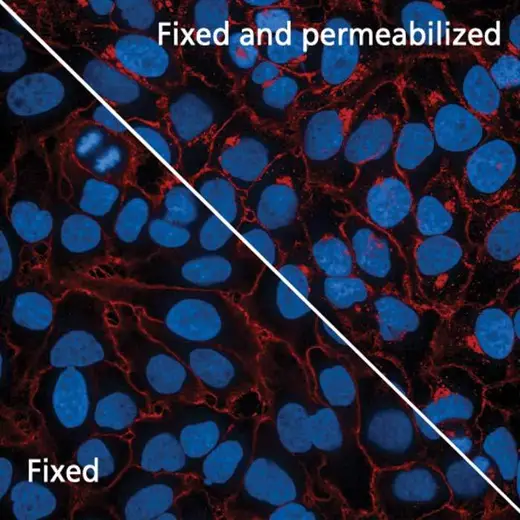
| Live cell staining | Yes |
| Fixed cell staining | Yes. See Product Information Sheet for more information |
| Equivalent number of microplates | 36-100 x 96-well microplates 30-100 x 384-well microplates 55-160 x 1536-well microplates |
Specifications
| Color | Red |
|---|
| Brand | PhenoVue™ |
|---|---|
| Detection Method | Fluorescence |
| Filter | Texas-Red |
| Fluorophore | PhenoVue™ Fluor 594 |
| Organelle and Cell Compartment | Golgi Apparatus |
| Product Compatibility | Live and fixed samples |
| Shipping Conditions | Shipped Ambient |
| Type | Individual Reagent |
| Unit Size | 5 vials |
Image gallery
Spectra Viewer
声明 :本官网所有报价均为常温或者蓝冰运输价格,如有产品需要干冰运输,需另外加收干冰运输费。

 危险品化学品经营许可证(不带存储) 许可证编号:沪(杨)应急管危经许[2022]202944(QY)
  营业执照(三证合一)