


 用小程序,查商品更便捷


Dot Blot
1:1000WB
1:1000ICC
1:100IP
1:50
Stat5, which stands for Signal Transducer and Activator of Transcription 5, is a transcription factor that plays a pivotal role in cellular signaling pathways. It is a key player in cellular signaling, particularly in response to cytokine receptors such as IL-2, IL-3, IL-5, and GM-CSF. Upon binding of these cytokines to their respective receptors, a cascade of signaling events is triggered, ultimately leading to the phosphorylation of Stat5 (e.g., at Tyr694). The phosphorylated Stat5 forms dimers and translocates to the nucleus, where it acts as a transcription factor to regulate the expression of target genes.
12 months from date of receipt / reconstitution, -20 °C as supplied
参考图片
Dot blot result of Phospho-Stat5 (Tyr694) Recombinant Rabbit mAb
Lane 1: Phospho-Stat5 (Tyr694) peptide
Lane 2: Stat5 unmodified peptide
Primary antibody: Phospho-Stat5 (Tyr694) Recombinant Rabbit mAb at 1/1000 dilution
Secondary antibody: Goat Anti-rabbit IgG, (H+L), HRP conjugated at 1/10000 dilution
WB result of Phospho-Stat5 (Tyr694) Recombinant Rabbit mAb
Primary antibody: Phospho-Stat5 (Tyr694) Recombinant Rabbit mAb at 1/1000 dilution
Lane 1: untreated A431 whole cell lysate 20 µg
Lane 2: A431 treated with 100 ng/ml EGF for 5 minutes whole cell lysate 20 µg
Secondary antibody: Goat Anti-rabbit IgG, (H+L), HRP conjugated at 1/10000 dilution
Predicted MW: 91 kDa
Observed MW: 90 kDa
ICC analysis of A431 cells treated with EGF (100ng/mL,5min) (top panel) and A431 cells untreated with EGF (100ng/mL,5min) (below panel). Anti-Phospho-Stat5 (Tyr694) antibody was used at 1/100 dilution (Green) and incubated overnight at 4°C. Goat polyclonal Antibody to Rabbit IgG - H&L (Alexa Fluor® 488) was used as secondary antibody at 1/1000 dilution. The cells were fixed with 100% ice-cold methanol and permeabilized with 0.1% PBS-Triton X-100. Nuclei were counterstained with DAPI (Blue). Counterstain with tubulin (Red).
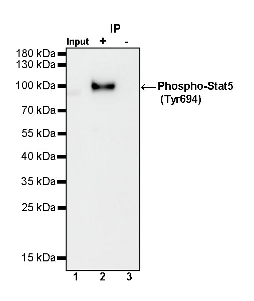
Phospho-Stat5 (Tyr694) Rabbit mAb at 1/50 dilution (1 µg) immunoprecipitating Phospho-Stat5 (Tyr694) in 0.4 mg A431 treated with 100 ng/ml EGF for 5 minutes whole cell lysate.
Western blot was performed on the immunoprecipitate using Phospho-Stat5 (Tyr694) Rabbit mAb at 1/1000 dilution.
Secondary antibody (HRP) for IP was used at 1/1000 dilution.
Lane 1: A431 treated with 100 ng/ml EGF for 5 minutes whole cell lysate 20 µg (Input)
Lane 2: Phospho-Stat5 (Tyr694) Rabbit mAb IP in A431 treated with 100 ng/ml EGF for 5 minutes whole cell lysate
Lane 3: Rabbit monoclonal IgG IP in A431 treated with 100 ng/ml EGF for 5 minutes whole cell lysate
Predicted MW: 91 kDa
Observed MW: 90 kDa

 危险品化学品经营许可证(不带存储) 许可证编号:沪(杨)应急管危经许[2022]202944(QY)
  营业执照(三证合一)