


 用小程序,查商品更便捷


Functional Assay
1:500FC
1:500
This antibody recognizes the same epitope as clone OKT3.
CD3 (cluster of differentiation 3) is a protein complex and T cell co-receptor that is involved in activating both the cytotoxic T cell (CD8+ naive T cells) and T helper cells (CD4+ naive T cells). It is composed of four distinct chains. In mammals, the complex contains a CD3γ chain, a CD3δ chain, and two CD3ε chains. These chains associate with the T-cell receptor (TCR) and the CD3-zeta (ζ-chain) to generate an activation signal in T lymphocytes. The TCR, CD3-zeta, and the other CD3 molecules together constitute the TCR complex. The CD3–T cell receptor (TCR) complex plays a central role in the T-cell-mediated immunoresponse as it is involved in the recognition of antigens and subsequent signal transduction and activation of immunocompetent T lymphocytes. Because CD3 is required for T cell activation, drugs (often monoclonal antibodies) that target it are being investigated as immunosuppressant therapies (e.g., otelixizumab, teplizumab) for type 1 diabetes and other autoimmune diseases.
12 months from date of receipt / reconstitution, 2 to 8 °C as supplied
参考图片
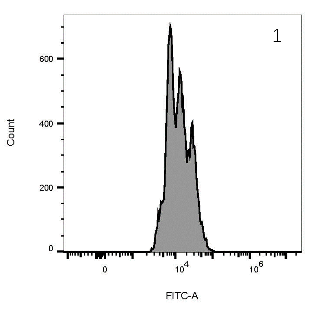
Flow cytometric analysis of Human peripheral blood mononuclear cells (PBMC) labeling CD3 with purified antibody at 1/500 dilution (0.1 μg) (Right) compared with a Rabbit monoclonal IgG isotype control (Left). Goat Anti-Mouse IgG Alexa Fluor® 488 was used as the secondary antibody.
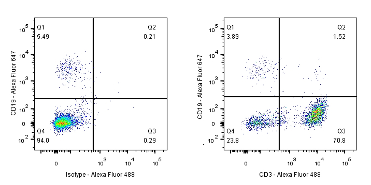
Cells were surface stained with CD19-Alexa Fluor® 647, then stained with rabbit IgG (Left) / anti-CD3 (Right) separately. CD3 and CD19 are mutually exclusive expressed in human PBMCs. Gated on total viable cells.
T cell proliferation Assay with CD3(S0B0009) &CD28 (S0B0010)
T cell proliferation Assay with CD3(S0B0009) &CD28 (S0B0010)
Procedure
1. Isolate human PBMCs according to the manufacturer’s instructions.
2. Suspend PBMCs in medium at 106 cells/mL.
3. Do one of the following depending on the format you are using:
· For the soluble format, incubate 106 cells with CD3 Mouse mAb (OKT3) at 1 µg/mL final concentration in culture for 3 days (5% CO2, 37°C).
· For the immobilized format, coat the CD3 Mouse mAb (OKT3) onto a plate at 5 to 10 μg/mL at 4°C, overnight. Add cells to the CD3 Mouse mAb (OKT3)-coated plate.
4. Wash the cells twice and resuspend them in medium at 106 cells/mL.
5. Distribute the cells in a round-bottom plate at 105 cells per well.
6. [Optional for co-stimulation] Add CD28 Mouse mAb (15E8) and optionally Protein G at 5 µg/mL each final concentration, and incubate the plate for an additional 3 days (5% CO2 at 37°C).
 危险品化学品经营许可证(不带存储) 许可证编号:沪(杨)应急管危经许[2022]202944(QY)
  营业执照(三证合一)