


 下载产品说明书 下载SDS
 用小程序,查商品更便捷


 收藏
收藏
 对比
对比 咨询
咨询Scientific Data
 View Larger
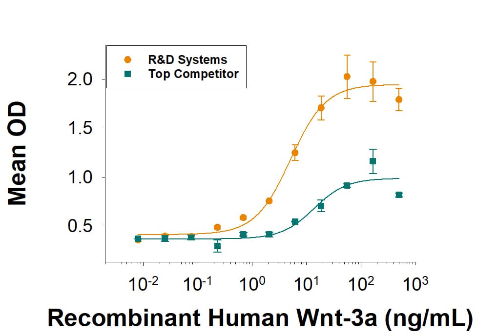
Recombinant Human Wnt-3a (Catalog # 5036-WN) induces alkaline phosphatase production by the MC3T3-E1 mouse preosteoblast cell line. The ED50 is 1.7-fold better with more than twice the maximum response compared to the top competitor's Wnt-3a.
 View Larger
The lot-to-lot consistency of Recombinant Human Wnt-3a Protein (Catalog # 5036-WN) was assessed by testing the ability of three independent lots of the protein to induce alkaline phosphatase production in the MC3T3-E1 mouse preosteoblast cell line. Each trace on the graph represents data obtained from Recombinant Human Wnt-3a from a different manufacturing run. The ED50 for this effect is 5.00-25.0 ng/mL.
 View Larger
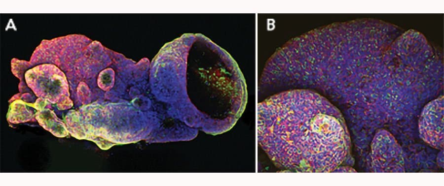
iPSC-derived human intestinal organoids were cultured using Cultrex™ UltiMatrix RGF Basement Membrane Extract (BME001-05) and intestinal organoid culture medium, which includes Recombinant Human Wnt-3a (Catalog # 5036-WN), Recombinant Human EGF (236-EG), Recombinant Human Noggin (6057-NG), and Recombinant Human R-Spondin 1 (4645-RS), along with the other reagents listed in the intestinal organoid culture medium recipe in the human intestinal organoid culture protocol. (A) Human intestinal organoids were stained using a Rat Anti-Human/Mouse/Rat Vimentin Monoclonal Antibody (MAB2105; green) and a Goat Anti-Human/Mouse Desmin Antigen Affinity-purified Polyclonal Antibody (AF3844; red) to visualize myofibroblast cells and counterstained with DAPI (5748; blue). (B) Human intestinal organoids were stained using a Goat Anti-Human/Mouse E-Cadherin Antigen Affinity-purified Polyclonal Antibody (AF748; green) and a Mouse Anti-Human MUC2 Monoclonal Antibody (Novus Biologicals, Catalog # NBP2-44431; red) and counterstained with DAPI (5748; blue).
 View Larger
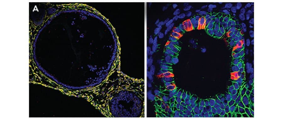
Adult stem cells isolated from human descending colon were embedded in Cultrex UltiMatrix RGF Basement Membrane Extract (BME001-05) and cultured for 30 days in intestinal organoid culture medium, which includes Recombinant Human Wnt-3a (Catalog # 5036-WN), Recombinant Human EGF (236-EG), Recombinant Human Noggin (6057-NG), and Recombinant Human R-Spondin 1 (4645-RS), along with the other reagents listed in the intestinal organoid culture medium recipe in the human intestinal organoid culture protocol. (A) Organoids were fixed and stained with a Mouse Anti-Human MUC2 Monoclonal Antibody (Novus Biologicals; Catalog # NBP2-44431; green) to visualize intestinal goblet cells and counterstained with a Goat Anti-Human/Mouse E-Cadherin Antigen Affinity-purified Polyclonal Antibody (AF748; red) and DAPI (5748; blue). The image shown was taken at 10x magnification. (B) Organoids were fixed and stained with a Mouse Anti-Human Chromogranin A Monoclonal Antibody (MAB90981; green) to visualize enteroendocrine cells and counterstained with a Goat Anti-Human/Mouse E-Cadherin Antigen Affinity-purified Polyclonal Antibody (AF748; red) and DAPI (5748; blue). The image shown was taken at 20x magnification.
Carrier Free
CF stands for Carrier Free (CF). We typically add Bovine Serum Albumin (BSA) as a carrier protein to our recombinant proteins. Adding a carrier protein enhances protein stability, increases shelf-life, and allows the recombinant protein to be stored at a more dilute concentration. The carrier free version does not contain BSA.
In general, we advise purchasing the recombinant protein with BSA for use in cell or tissue culture, or as an ELISA standard. In contrast, the carrier free protein is recommended for applications, in which the presence of BSA could interfere.
5036-WN
| Formulation | Lyophilized from a 0.2 μm filtered solution in PBS, EDTA and CHAPS with BSA as a carrier protein. |
| Reconstitution | Reconstitute at 200 μg/mL in sterile PBS containing at least 0.1% human or bovine serum albumin. |
| Shipping | The product is shipped at ambient temperature. Upon receipt, store it immediately at the temperature recommended below. |
| Stability & Storage: | Use a manual defrost freezer and avoid repeated freeze-thaw cycles.
|
5036-WN/CF
| Formulation | Lyophilized from a 0.2 μm filtered solution in PBS, EDTA and CHAPS. |
| Reconstitution | Reconstitute at 200 μg/mL in sterile PBS. |
| Shipping | The product is shipped at ambient temperature. Upon receipt, store it immediately at the temperature recommended below. |
| Stability & Storage: | Use a manual defrost freezer and avoid repeated freeze-thaw cycles.
|
Recombinant Human Wnt-3a Protein Summary
Product Specifications
Optimal concentrations should be determined by each laboratory for each application.
Ser19-Lys352
Analysis

Background: Wnt-3a
Wnt-3a is one of 19 vertebrate members of the Wingless-type MMTV integration site (Wnt) family of highly conserved cysteine-rich secreted glycoproteins important for normal developmental processes (1). Wnts bind to the cell surface Frizzled family receptors in conjunction with low-density lipoprotein receptor-related protein family receptors (LRP5 or 6) resulting in the stabilization of intracellular beta -catenin levels (2). As intracellular beta -catenin levels rise, beta -catenin binds to TCF/LEF transcription factors leading to expression of Wnt target genes (3). Endo-IWR 1 (Catalog # 3532, # PSM1324) is a cell-permeant small molecule inhibitor of Axin turnover that suppresses Wnt signal transduction by stabilizing the beta -catenin destruction complex (4). Wnt-3a is a 44 kDa secreted hydrophobic glycoprotein containing a conserved pattern of 24 cysteine residues (5). Wnt-3a has two N-linked glycosylation sites (Asn 87, Asn 298), and Ser 209 is modified with palmitoleic acid (6). Glycosylation and acylation are essential for efficient Wnt secretion and biological activity, respectively (6, 7). Human Wnt-3a shares 96% amino acid (aa) identity with mouse mouse, bovine and canine Wnt-3a, and 89%, 86% and 84% aa identity with chicken, Xenopus and zebrafish Wnt-3a, respectively. It also shares 87% aa identity with Wnt3. During embryonic development, Wnt-3a is necessary for proper development of the hippocampus, anterior-posterior patterning, somite development, and tailbud formation (9-12). Wnt-3a also promotes self-renewal of hematopoietic stem cells, neural stem cells, and embryonic stem cells (13, 14).
- Willert, K. and Nusse, R. (2012) Cold Spring Harb. Perspect. Biol. 4:a007864.
- MacDonald, B.T. and X. He (2012) Cold Spring Harb. Perspect. Biol. 4:a007880.
- Korinek, V. et al. (1997) Science 275:1784.
- Chen, B. et al. (2009) Nat. Chem. Biol. 5:100.
- Smolich, B.D. et al. (1993) Mol. Biol. Cell 4:1267.
- Takada, R. et al. (2006) Dev. Cell 11:791.
- Komekado, H. (2007) Genes Cells 12:521.
- Dunty Jr. W. C. et al. (2008) Development 135:85.
- Ikeya, M. and S. Takada (2001) Mech. Dev. 103:27.
- Lee, S. M. et al. (2000) Development 127:457.
- Takada, S. et al. (1994) Genes Dev. 8:174.
- Willert, K. et al. (2003) Nature 423:6938.
- Kalani, M.Y. et al. (2008) Proc. Natl. Acad. Sci. USA 105:16970.
- Ten Berge, D. et al. (2011) Nat. Cell Biol. 13:1070.

 危险品化学品经营许可证(不带存储) 许可证编号:沪(杨)应急管危经许[2022]202944(QY)
  营业执照(三证合一)