


BD Horizon™ BV421 Mouse Anti-Human CD4


 下载产品说明书 下载SDS
 用小程序,查商品更便捷


 收藏
收藏
 对比
对比 咨询
咨询

参考图片
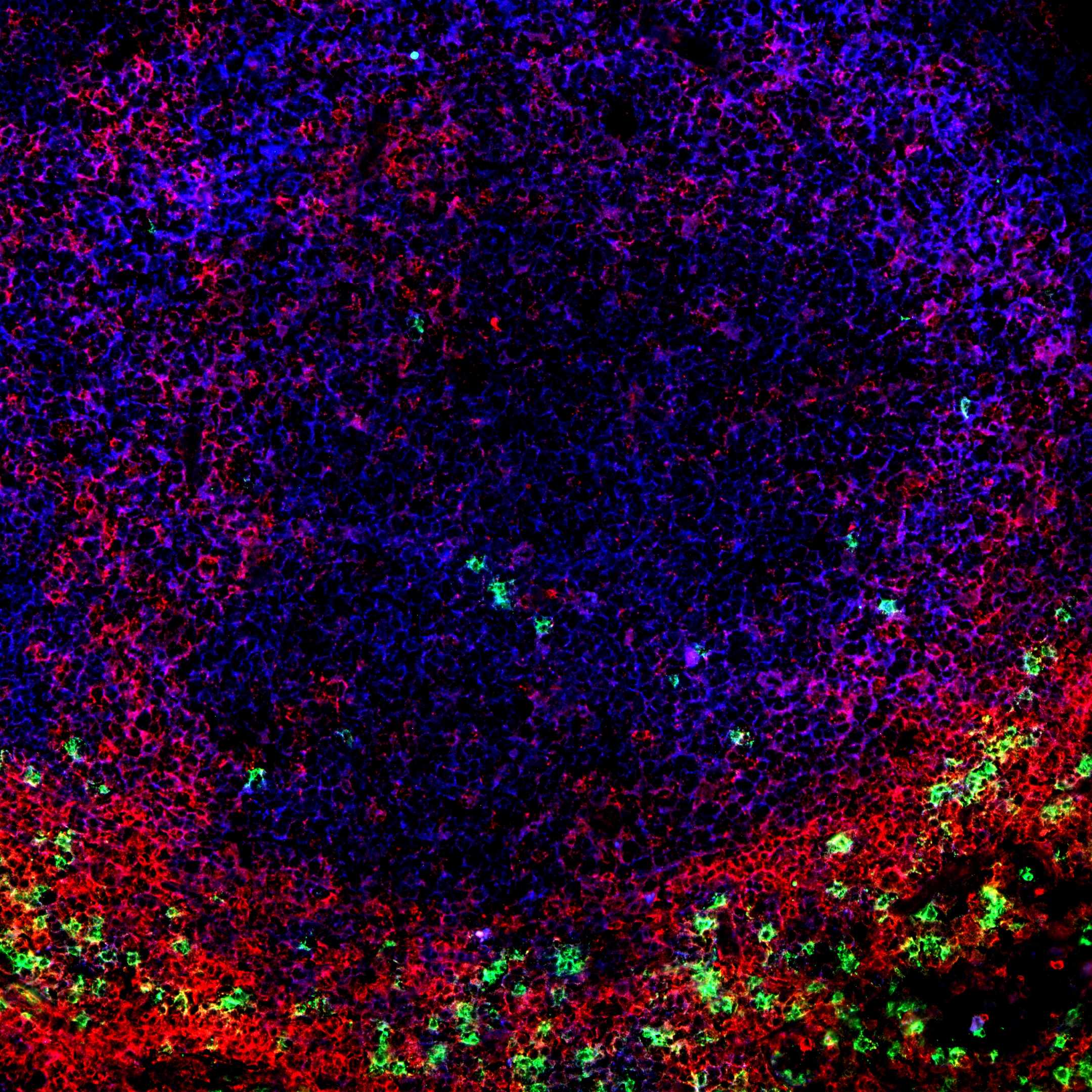
Immunohistofluorescent analysis of CD4 expression by cells within human tonsil. A human tonsil cryosection (5 µm) was fixed with BD Cytofix™ Fixation Buffer (Cat. No. 554655), blocked with 5% goat serum and 1% BSA diluted in 1x PBS, and stained with BD Pharmingen™ Purified Mouse Anti-Human CD8 antibody (Cat. No. 555364) followed by BD Horizon™ BV480 Goat Anti-Mouse Ig second step antibody (Cat. No. 564877, pseudo-colored green). Sections were thoroughly washed, then stained with BD Horizon™ BV421 Mouse Anti-Human CD4 antibody (Cat. No. 562424/562425, pseudo-colored red) and Alexa Fluor® 488 Mouse Anti-Human CD19 antibody (Cat. No. 557697, pseudo-colored blue). Images were captured on a standard epifluorescence microscope. Original magnification, 20x.
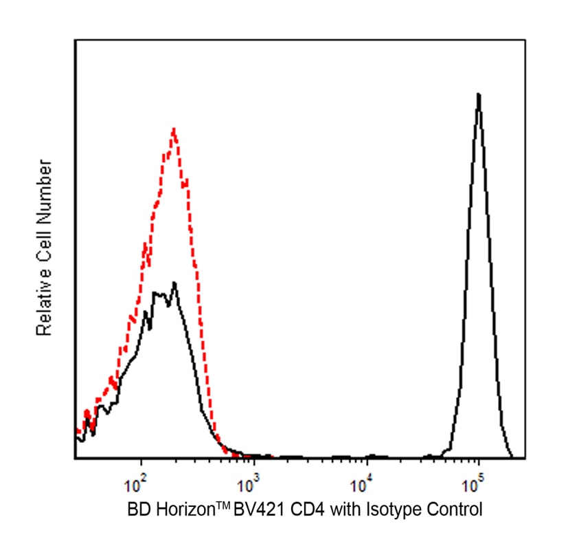
Flow cytometric analysis of CD4 expression on human peripheral blood lymphocytes. Human whole blood was stained at 4°C with the BD Horizon™ BV421 Mouse anti-Human CD4 antibody (Cat. No. 562424/562425; solid line histogram) or with a BD Horizon™ BV421 Mouse IgG1, κ Isotype Control (Cat. No. 562438; dashed line histogram). The erythrocytes were lysed with BD Pharm Lyse™ Lysing Buffer (Cat. No. 555899). The fluorescence histograms were derived from events with the forward and side light-scatter characteristics of viable lymphocytes. Flow cytometry was performed using a BD FACSCanto™ II Flow Cytometer System.
Immunohistofluorescent analysis of CD4 expression by cells within human tonsil. A human tonsil cryosection (5 µm) was fixed with BD Cytofix™ Fixation Buffer (Cat. No. 554655), blocked with 5% goat serum and 1% BSA diluted in 1x PBS, and stained with BD Pharmingen™ Purified Mouse Anti-Human CD8 antibody (Cat. No. 555364) followed by BD Horizon™ BV480 Goat Anti-Mouse Ig second step antibody (Cat. No. 564877, pseudo-colored green). Sections were thoroughly washed, then stained with BD Horizon™ BV421 Mouse Anti-Human CD4 antibody (Cat. No. 562424/562425, pseudo-colored red) and Alexa Fluor® 488 Mouse Anti-Human CD19 antibody (Cat. No. 557697, pseudo-colored blue). Images were captured on a standard epifluorescence microscope. Original magnification, 20x.

 危险品化学品经营许可证(不带存储) 许可证编号:沪(杨)应急管危经许[2022]202944(QY)
  营业执照(三证合一)