


1/2

品牌: BD Pharmingen
 下载产品说明书
 用小程序,查商品更便捷


 收藏
收藏
 对比
对比 咨询
咨询反应种属:
Human (QC Testing), Rhesus, Cynomolgus, Baboon (Tested in Development)
Human (QC Testing), Rhesus, Cynomolgus, Baboon (Tested in Development)
来源宿主:
Mouse BALB/c IgG1, κ
Mouse BALB/c IgG1, κ
产品介绍
产品信息
耦联标记
APC-Cy7

抗原名称
CD8
宿主
Mouse BALB/c IgG1, κ
免疫原
Human Peripheral Blood T Cells
简单描述
The SK1 monoclonal antibody specifically binds to CD8 alpha (CD8α). CD8α is a type I transmembrane glycoprotein and a member of the immunoglobulin superfamily. CD8α is expressed by the majority of thymocytes, by subpopulations of αβ T cells and γδ T cells and by some NK cells. Cell surface CD8α is expressed either as a disulfide-linked homodimer (CD8αα) or as a heterodimer (CD8αβ) when disulfide-bonded to a CD8 beta chain (CD8β). CD8-positive αβ T cells coexpress both CD8αα homodimers and CD8αβ heterodimers whereas som
e γδ T cells and NK cells express CD8αα homodimers. CD8 plays important roles in T cell activation and selection. The extracellular IgSF domain of CD8α binds to a non-polymorphic determinant on HLA class I molecules (α3 domain) and enables CD8 to function as a co-receptor with MHC class I-restricted TCR during T cell recognition of antigen. The cytoplasmic domain of CD8α associates with Lck, a Src family protein tyrosine kinase that is involved in intracellular signaling.
商品描述
The SK1 monoclonal antibody specifically binds to CD8 alpha (CD8α). CD8α is a type I transmembrane glycoprotein and a member of the immunoglobulin superfamily. CD8α is expressed by the majority of thymocytes, by subpopulations of αβ T cells and γδ T cells and by some NK cells. Cell surface CD8α is expressed either as a disulfide-linked homodimer (CD8αα) or as a heterodimer (CD8αβ) when disulfide-bonded to a CD8 beta chain (CD8β). CD8-positive αβ T cells coexpress both CD8αα homodimers and CD8αβ heterodimers whereas som
e γδ T cells and NK cells express CD8αα homodimers. CD8 plays important roles in T cell activation and selection. The extracellular IgSF domain of CD8α binds to a non-polymorphic determinant on HLA class I molecules (α3 domain) and enables CD8 to function as a co-receptor with MHC class I-restricted TCR during T cell recognition of antigen. The cytoplasmic domain of CD8α associates with Lck, a Src family protein tyrosine kinase that is involved in intracellular signaling.
同种型
Mouse BALB/c IgG1, κ
克隆号
SK1
产品详情
APC-Cy7
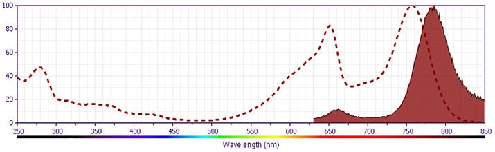
APC-Cy7 dye is a part of the BD APC red family of dyes. This tandem fluorochrome is comprised of a Allophycocyanin (APC) donor that has excitation maxima (Ex Max) of 651 nm and an acceptor dye, Cy™7, with an emission maximum (Em Max) at 779 nm. APC-Cy7 can be excited by the Red (627-640 nm) laser and detected using an optical filter centered near 780 nm (e.g., a 760/60 nm bandpass filter). Please ensure that your instrument’s configurations (lasers and optical filters) are appropriate for this dye.
APC-Cy7
Red 627-640 nm
651 nm
779 nm
应用
实验应用
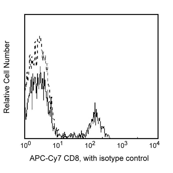
Flow cytometry (Routinely Tested)
推荐用量
5 µl
反应种属
Human (QC Testing), Rhesus, Cynomolgus, Baboon (Tested in Development)
背景
别名
CD8α; CD8A; CD8 alpha; Leu2a; MAL; T8; p32
制备和贮存
存储溶液
Aqueous buffered solution containing BSA and ≤0.09% sodium azide.
保存方式
Aqueous buffered solution containing BSA and ≤0.09% sodium azide.
文献
文献
研发参考(13)
1. Beavis AJ, Pennline KJ. Allo-7: a new fluorescent tandem dye for use in flow cytometry. Cytometry. 1996; 24(4):390-395. (Biology).
2. Bernard A, Boumsell L, Hill C. Joint report of the first international workshop on human leucocyte differentiation antigens by the investigators of the participating laboratories: T2 protocol. In: Bernard A. A. Bernard .. et al., ed. Leucocyte typing : human leucocyte differentiation antigens detected by monoclonal antibodies : specification, classification, nomenclature = Typage leucocytaire : antigènes de différenciation leucocytaire humains révélés par les anticorps monoclonaux : "Rapports des études communes". Berlin New York: Springer-Verlag; 1984:25-60.
3. Dongworth DW, Gotch FM, Carter NP, Hildreth PDK, McMichael AJ. Inhibition of virus-specific, HLA-restricted, T cell-mediated lysis by monoclonal anti-T cell antibodies. In: Bernard A. A. Bernard .. et al., ed. Leucocyte typing : human leucocyte differentiation antigens detected by monoclonal antibodies : specification, classification, nomenclature = Typage leucocytaire : antigènes de différenciation leucocytaire humains révélés par les anticorps monoclonaux : "Rapports des études communes". Berlin New York: Springer-Verlag; 1984:320-328.
4. Engleman EG, Benike CJ, Glickman E, Evans RL. Antibodies to membrane structures that distinguish suppressor/cytotoxic and helper T lymphocyte subpopulations block the mixed leukocyte reaction in man. J Exp Med. 1981; 154(1):193-198. (Clone-specific: Cell separation, Flow cytometry, Functional assay, Inhibition).
5. Engleman EG, Benike CJ, Grumet FC, Evans RL. Activation of human T lymphocyte subsets: helper and suppressor/cytotoxic T cells recognize and respond to distinct histocompatibility antigens. J Immunol. 1981; 127(5):2124-2129. (Clone-specific: Cell separation, Flow cytometry, Fluorescence activated cell sorting).
6. Evans RL, Wall DW, Platsoucas CD, et al. Thymus-dependent membrane antigens in man: inhibition of cell-mediated lympholysis by monoclonal antibodies to TH2 antigen. Proc Natl Acad Sci U S A. 1981; 78(1):544-548. (Immunogen: Flow cytometry, Functional assay, Inhibition).
7. Jonker M, Meurs G. Monoclonal antibodies specific for B cells, cytotoxic/suppressor T cells, and a subset of cytotoxic/suppressor T cells in the Rhesus monkey. In: Bernard A. A. Bernard .. et al., ed. Leucocyte typing : human leucocyte differentiation antigens detected by monoclonal antibodies : specification, classification, nomenclature = Typage leucocytaire : antigènes de différenciation leucocytaire humains révélés par les anticorps monoclonaux : "Rapports des études communes". Berlin New York: Springer-Verlag; 1984:328-336.
8. Knapp W. W. Knapp .. et al., ed. Leucocyte typing IV : white cell differentiation antigens. Oxford New York: Oxford University Press; 1989:1-1182.
9. Ledbetter JA, Evans RL, Lipinski M, Cunningham-Rundles C, Good RA, Herzenberg LA. Evolutionary conservation of surface molecules that distinguish T lymphocyte helper/inducer and cytotoxic/suppressor subpopulations in mouse and man. J Exp Med. 1981; 153(2):310-323. (Clone-specific: Flow cytometry, Immunoprecipitation).
10. McMichael AJ. A.J. McMichael .. et al., ed. Leucocyte typing III : white cell differentiation antigens. Oxford New York: Oxford University Press; 1987:1-1050.
11. Roederer M, Kantor AB, Parks DR, Herzenberg LA. Cy7PE and Cy7APC: bright new probes for immunofluorescence. Cytometry. 1996; 24(3):191-197. (Biology).
12. Schlossman SF. Stuart F. Schlossman .. et al., ed. Leucocyte typing V : white cell differentiation antigens : proceedings of the fifth international workshop and conference held in Boston, USA, 3-7 November, 1993. Oxford: Oxford University Press; 1995.
13. Warner NL, Lanier LL, Jackson A, Babcock G, Evans R. Multiparameter approaches to FACS analysis of human leucocyte cell surface antigens. In: Bernard A. A. Bernard .. et al., ed. Leucocyte typing : human leucocyte differentiation antigens detected by monoclonal antibodies. Berlin New York: Springer-Verlag; 1984:621-630.
数据库链接
Entrez-Gene ID
925
参考图片
Profile of peripheral blood lymphocytes analyzed by flow cytometry
声明 :本官网所有报价均为常温或者蓝冰运输价格,如有产品需要干冰运输,需另外加收干冰运输费。

 危险品化学品经营许可证(不带存储) 许可证编号:沪(杨)应急管危经许[2022]202944(QY)
  营业执照(三证合一)