


BD Horizon™ BV421 Rat Anti-Mouse F4/80


 下载产品说明书 下载SDS
 用小程序,查商品更便捷


 收藏
收藏
 对比
对比 咨询
咨询

参考图片
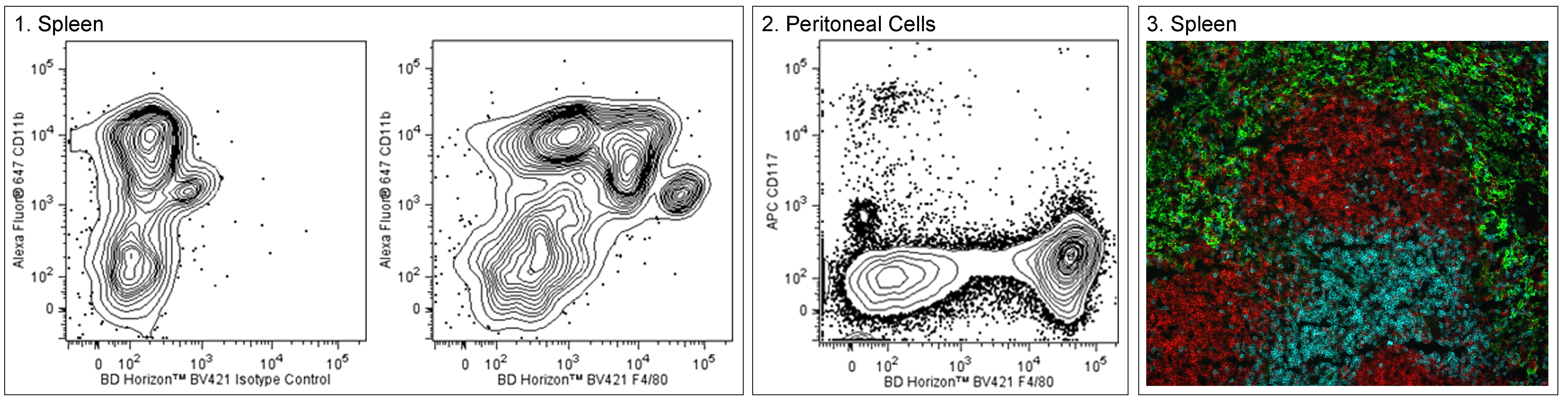
Two-color flow cytometric analysis of mouse F4/80 expression. Panel 1 - Spleen. C57BL/6 mouse splenic leucocytes were preincubated with Purified Rat Anti-Mouse CD16/CD32 antibody (Mouse BD Fc Block™) (Cat. No. 553141/553142) and stained with Alexa Fluor® 647 Rat Anti-Mouse CD11b antibody (Cat. No. 557686) and either BD Horizon™ BV421 Rat IgG2a Isotype Control (Cat. No. 562602; Left Plot) or BD Horizon BV421 Rat Anti-Mouse F4/80 antibody (Cat. No. 565411; Right Plot). The two-color flow cytometric contour plot showing the correlated expression of F4/80 (or Ig Isotype control staining) versus CD11b was derived from gated events with the forward and side light-scatter characteristics of viable monocytes. Panel 2 - Peritoneal. C57BL/6 mouse peritoneal cells were preincubated with Mouse BD Fc Block™ and stained with APC Rat Anti-Mouse CD117 (Cat. No. 553356/561074) and BV421 Rat Anti-Mouse F4/80 antibodies. The two-color contour plot showing the correlated expression of F4/80 versus CD117 was derived from gated events with the light-scatter characteristics of viable peritoneal cells. Flow cytometric analyses were performed using a BD LSRFortessa™ Cell Analyzer System. Image analysis of F4/80 expression in mouse spleen. Panel 3 - Spleen. A section from a paraformaldehyde-fixed and frozen normal C57BL/6 mouse spleen was stained with BV421 Rat Anti-Mouse F4/80 (pseudo-colored green), Alexa Fluor® 647 Rat Anti-Mouse CD45R/B220 (Cat. No. 557683; pseudo-colored red), and BD Horizon™ BV480 Anti-CD3 (pseudo-colored aqua) antibodies. The image was captured on a standard Epifluorescence microscope using 392/23nm excitation and 430/24nm emission.

 危险品化学品经营许可证(不带存储) 许可证编号:沪(杨)应急管危经许[2022]202944(QY)
  营业执照(三证合一)