


 下载产品说明书
 用小程序,查商品更便捷


 收藏
收藏
 对比
对比 咨询
咨询Disclaimer note: The observed molecular weight of the protein may vary from the listed predicted molecular weight due to post translational modifications, post translation cleavages, relative charges, and other experimental factors.
Disclaimer note: The observed molecular weight of the protein may vary from the listed predicted molecular weight due to post translational modifications, post translation cleavages, relative charges, and other experimental factors.

Expressed in a defined zone of basal keratinocytes in the deep outer root sheath of hair follicles. Also observed in sweat gland and mammary gland ductal and secretory cells, bile ducts, gastrointestinal tract, bladder urothelium, oral epithelia, esophagus, ectocervical epithelium (at protein level). Expressed in epidermal basal cells, in nipple epidermis and a defined region of the hair follicle. Also seen in a subset of vascular wall cells in both the veins and artery of human umbilical cord, and in umbilical cord vascular smooth muscle. Observed in muscle fibers accumulating in the costameres of myoplasm at the sarcolemma in structures that contain dystrophin and spectrin
Disclaimer note: The observed molecular weight of the protein may vary from the listed predicted molecular weight due to post translational modifications, post translation cleavages, relative charges, and other experimental factors.
参考图片
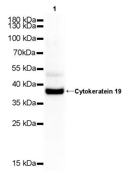
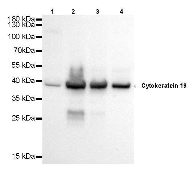
WB result of Cytokeratin 19 Rabbit mAb Primary antibody: Cytokeratin 19 Rabbit mAb at 1/500 dilution Lane 1: Hela whole cell lysate 20 µg Lane 2: MCF7 whole cell lysate 20 µg Lane 3: HT-29 whole cell lysate 20 µg Lane 4: HepG2 whole cell lysate 20 µgSecondary antibody: #abs20040 at 1/10000 dilution Predicted MW: 41 kDa Observed MW: 39 kDa Exposure time: 20s
WB result of Cytokeratin 19 Rabbit mAb Primary antibody: Cytokeratin 19 Rabbit mAb at 1/500 dilution Lane 1: mouse kidney lysate 20 µg Secondary antibody: #abs20040 at 1/10000 dilution Predicted MW: 41 kDa Observed MW: 39 kDa Exposure time: 120 s
WB result of Cytokeratin 19 Rabbit mAb Primary antibody: Cytokeratin 19 Rabbit mAb at 1/500 dilution Lane 1: rat kidney whole cell lysate 20 µg Secondary antibody: #abs20040 at 1/10000 dilution Predicted MW: 41 kDa Observed MW: 39 kDa Exposure time: 120 s
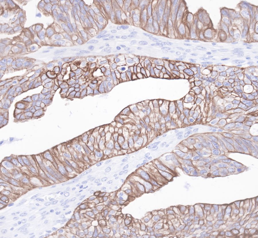
IHC shows positive staining in paraffin-embedded human colon. Anti-Cytokeratin19 antibody was used at 1/1000 dilution, Secondary antibody: #abs20040. Counterstained with hematoxylin. Heat mediated antigen retrieval with Tris/EDTA buffer pH9.0 was performed before commencing with IHC staining protocol.
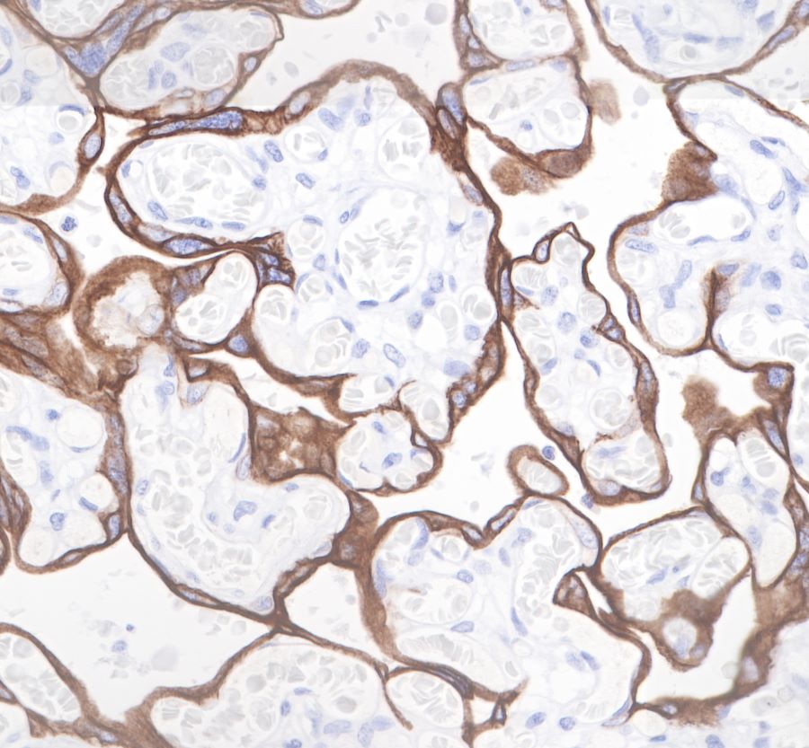
IHC shows positive staining in paraffin-embedded human placenta. Anti-Cytokeratin19 antibody was used at 1/1000 dilution, Secondary antibody: #abs20040. Counterstained with hematoxylin. Heat mediated antigen retrieval with Tris/EDTA buffer pH9.0 was performed before commencing with IHC staining protocol.
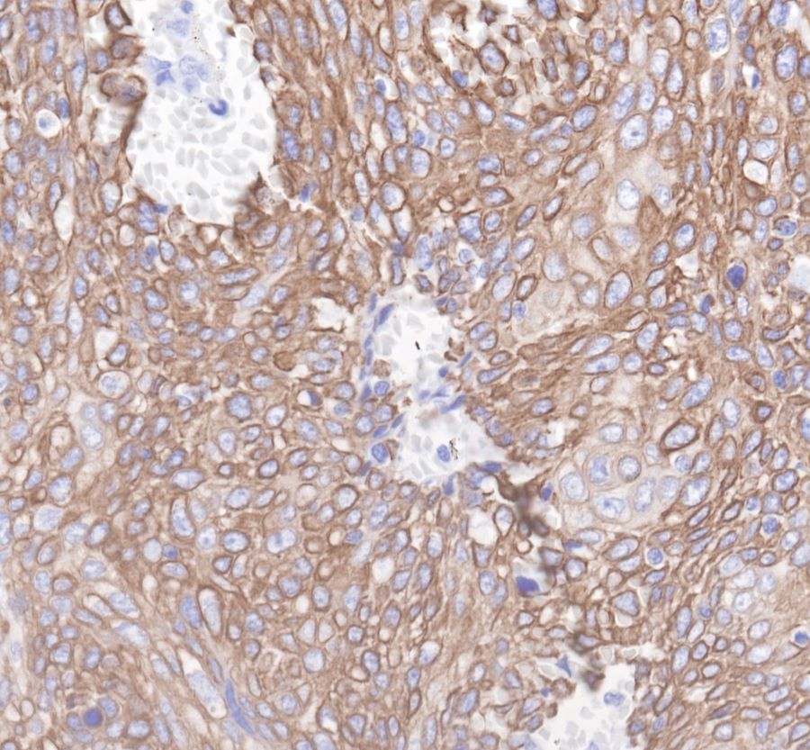
IHC shows positive staining in paraffin-embedded human ovarian cancer. Anti-Cytokeratin19 antibody was used at 1/1000 dilution, Secondary antibody: #abs20040. Counterstained with hematoxylin. Heat mediated antigen retrieval with Tris/EDTA buffer pH9.0 was performed before commencing with IHC staining protocol.
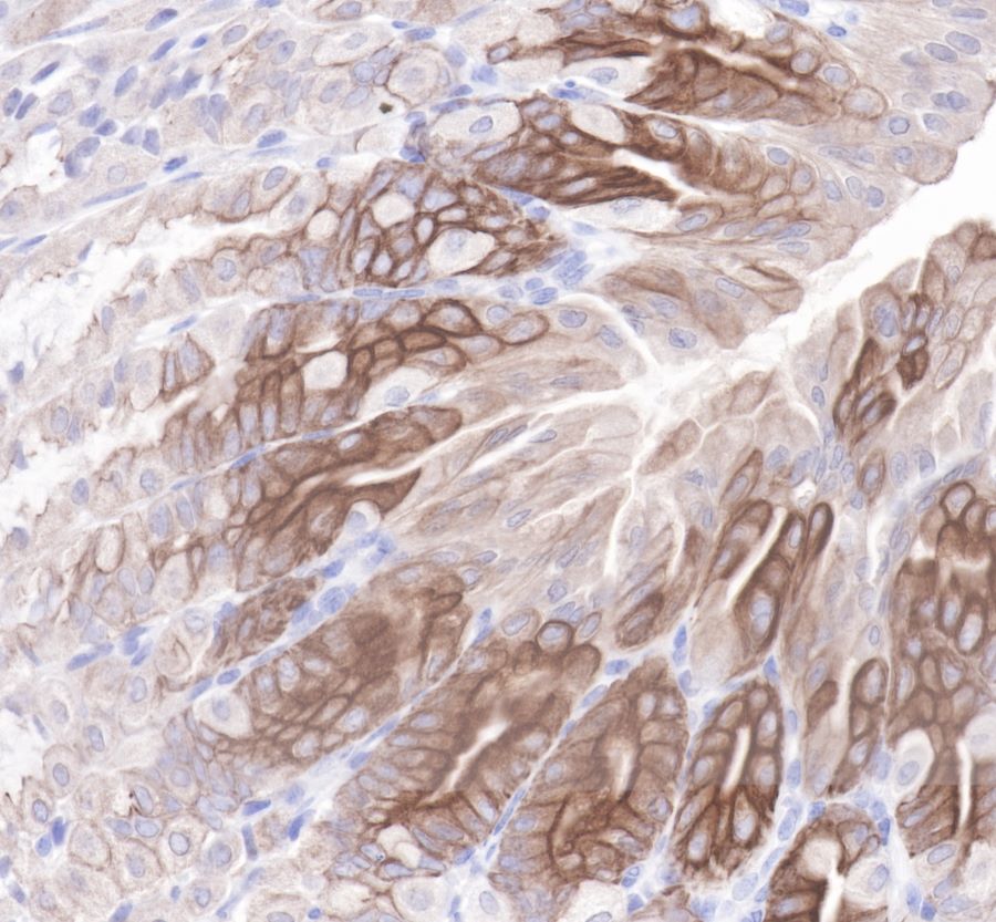
IHC shows positive staining in paraffin-embedded human cervix cancer. Anti-Cytokeratin19 antibody was used at 1/1000 dilution, Secondary antibody: #abs20040. Counterstained with hematoxylin. Heat mediated antigen retrieval with Tris/EDTA buffer pH9.0 was performed before commencing with IHC staining protocol.
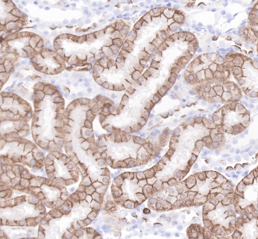
IHC shows positive staining in paraffin-embedded mouse stomach. Anti-Cytokeratin19 antibody was used at 1/1000 dilution, Secondary antibody: #abs20040. Counterstained with hematoxylin. Heat mediated antigen retrieval with Tris/EDTA buffer pH9.0 was performed before commencing with IHC staining protocol.
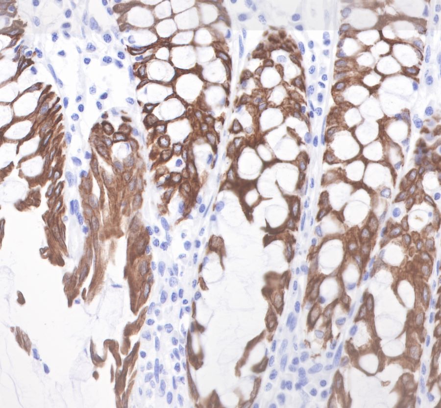
IHC shows positive staining in paraffin-embedded rat kidney. Anti-Cytokeratin19 antibody was used at 1/1000 dilution, Secondary antibody: #abs20040. Counterstained with hematoxylin. Heat mediated antigen retrieval with Tris/EDTA buffer pH9.0 was performed before commencing with IHC staining protocol.
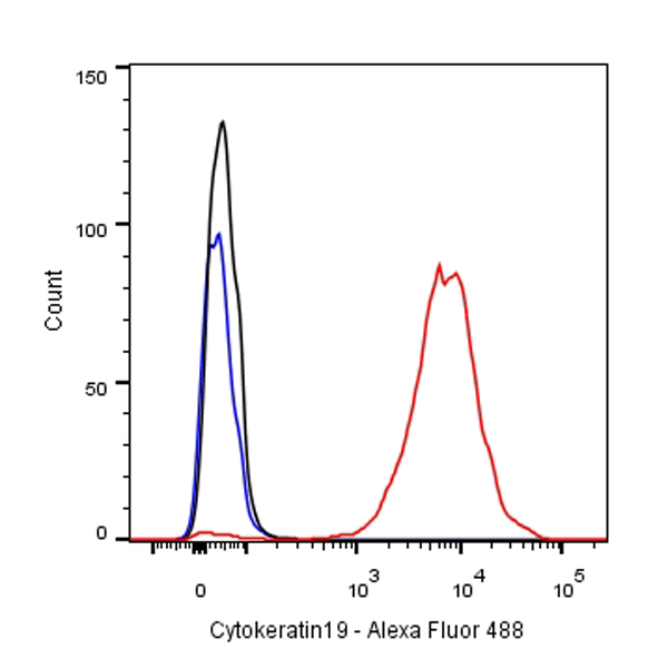
Flow cytometric analysis of MCF7 cells labelling Cytokeratin 19 antibody at 1/500 dilution/ (red) compared with a Rabbit monoclonal IgG (Black) isotype control and an unlabelled control (cells without incubation with primary antibody and secondary antibody) (Blue). Secondary antibody: #abs20040 at 1/1000 dilution was used as the secondary antibody.
Flow cytometric analysis of MCF7 cells labelling Cytokeratin 19 antibody at 1/500 dilution/ (red) compared with a Rabbit monoclonal IgG (Black) isotype control and an unlabelled control (cells without incubation with primary antibody and secondary antibody) (Blue). Goat Anti-Rabbit IgG Alexa Fluor® 488 at 1/1000 dilution was used as the secondary antibody.
IHC shows positive staining in paraffin-embedded human colon.
Anti-Cytokeratin19 antibody was used at 1/1000 dilution, followed by a Goat Anti-Rabbit IgG H&L (HRP) ready to use.
Counterstained with hematoxylin.
Heat mediated antigen retrieval with Tris/EDTA buffer pH9.0 was performed before commencing with IHC staining protocol.
IHC shows positive staining in paraffin-embedded human placenta.
Anti-Cytokeratin19 antibody was used at 1/1000 dilution, followed by a Goat Anti-Rabbit IgG H&L (HRP) ready to use.
Counterstained with hematoxylin.
Heat mediated antigen retrieval with Tris/EDTA buffer pH9.0 was performed before commencing with IHC staining protocol.
Anti-Cytokeratin19 antibody was used at 1/1000 dilution, followed by a Goat Anti-Rabbit IgG H&L (HRP) ready to use.
Counterstained with hematoxylin.
Heat mediated antigen retrieval with Tris/EDTA buffer pH9.0 was performed before commencing with IHC staining protocol.
IHC shows positive staining in paraffin-embedded human cervix cancer. Anti-Cytokeratin19 antibody was used at 1/1000 dilution, followed by a Goat Anti-Rabbit IgG H&L (HRP) ready to use.
Counterstained with hematoxylin.
Heat mediated antigen retrieval with Tris/EDTA buffer pH9.0 was performed before commencing with IHC staining protocol.
IHC shows positive staining in paraffin-embedded mouse stomach.
Anti-Cytokeratin19 antibody was used at 1/1000 dilution, followed by a Goat Anti-Rabbit IgG H&L (HRP) ready to use.
Counterstained with hematoxylin.
Heat mediated antigen retrieval with Tris/EDTA buffer pH9.0 was performed before commencing with IHC staining protocol.
IHC shows positive staining in paraffin-embedded rat kidney.
Anti-Cytokeratin19 antibody was used at 1/1000 dilution, followed by a Goat Anti-Rabbit IgG H&L (HRP) ready to use.
Counterstained with hematoxylin.
Heat mediated antigen retrieval with Tris/EDTA buffer pH9.0 was performed before commencing with IHC staining protocol.
WB resuLt of Cytokeratin 19 Rabbit mAb
Primary antibody: Cytokeratin 19 Rabbit mAb at 1/500 dilution
Lane 1: Hela whole cell lysate 20 µg
Lane 2: MCF7 whole cell lysate 20 µg
Lane 3: HT-29 whole cell lysate 20 µg
Lane 4: HepG2 whole cell lysate 20 µg
Secondary antibody: Goat Anti-Rabbit IgG, (H+L), HRP conjugated at 1/10000 dilution
Predicted MW: 41 kDa
Observed MW: 39 kDa
Exposure time: 20s
WB resuLt of Cytokeratin 19 Rabbit mAb
Primary antibody: Cytokeratin 19 Rabbit mAb at 1/500 dilution
Lane 1: mouse kidney lysate 20 µg
Secondary antibody: Goat Anti-Rabbit IgG, (H+L), HRP conjugated at 1/10000 dilution
Predicted MW: 41 kDa
Observed MW: 39 kDa
Exposure time: 120 s
WB resuLt of Cytokeratin 19 Rabbit mAb
Primary antibody: Cytokeratin 19 Rabbit mAb at 1/500 dilution
Lane 1: rat kidney whole cell lysate 20 µg
Secondary antibody: Goat Anti-Rabbit IgG, (H+L), HRP conjugated at 1/10000 dilution
Predicted MW: 41 kDa
Observed MW: 39 kDa
Exposure time: 120 s
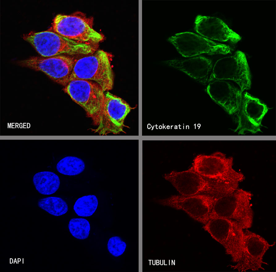
ICC shows positive staining in MCF7 cells. Anti-Cytokeratin 19 antibody was used at 1/250 dilution (Green) and incubated overnight at 4°C. Goat polyclonal Antibody to Rabbit IgG - H&L (Alexa Fluor® 488) was used as secondary antibody at 1/1000 dilution. The cells were fixed with 4% PFA and permeabilized with 0.1% PBS-Triton X-100. Nuclei were counterstained with DAPI (Blue). Counterstain with tubuLin (red).

 危险品化学品经营许可证(不带存储) 许可证编号:沪(杨)应急管危经许[2022]202944(QY)
  营业执照(三证合一)