


Rabbit anti-GPX4 Polyclonal Antibody


 下载产品说明书
 用小程序,查商品更便捷


 收藏
收藏
 对比
对比 咨询
咨询Disclaimer note: The observed molecular weight of the protein may vary from the listed predicted molecular weight due to post translational modifications, post translation cleavages, relative charges, and other experimental factors.
Disclaimer note: The observed molecular weight of the protein may vary from the listed predicted molecular weight due to post translational modifications, post translation cleavages, relative charges, and other experimental factors.

Glutathione peroxidase catalyzes the reduction of hydrogen peroxide, organic hydroperoxide, and lipid peroxides by reduced glutathione and functions in the protection of cells against oxidative damage. Human plasma glutathione peroxidase has been shown to be a selenium-containing enzyme and the UGA codon is translated into a selenocysteine. Through alternative splicing and transcription initiation, rat produces proteins that localize to the nucleus, mitochondrion, and cytoplasm. In humans, experimental evidence for alternative splicing exists; alternative transcription initiation and the cleavage sites of the mitochondrial and nuclear transit peptides need to be experimentally verified. [provided by RefSeq, Jul 2008]
Disclaimer note: The observed molecular weight of the protein may vary from the listed predicted molecular weight due to post translational modifications, post translation cleavages, relative charges, and other experimental factors.
参考图片
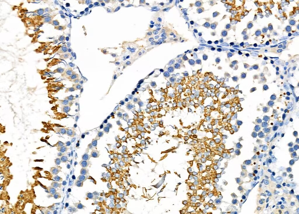
Rabbit anti-GPX4 Polyclonal Antibody at 1/100 staining Rat testis tissue by IHC-P. The sample was formaldehyde fixed and a heat mediated antigen retrieval step in citrate buffer was performed. The sample was then blocked and incubated with the primary antibody at 4°C overnight. An HRP conjugated anti-Rabbit antibody was used as the secondary antibody.
Western blot analysis of extracts from various samples, using GPX4 antibody at 1/1000 dilution. Lane 1: Mouse testis. Lane 2: Ec304 cells(heat-shock treatment). Observed bands:20kD.
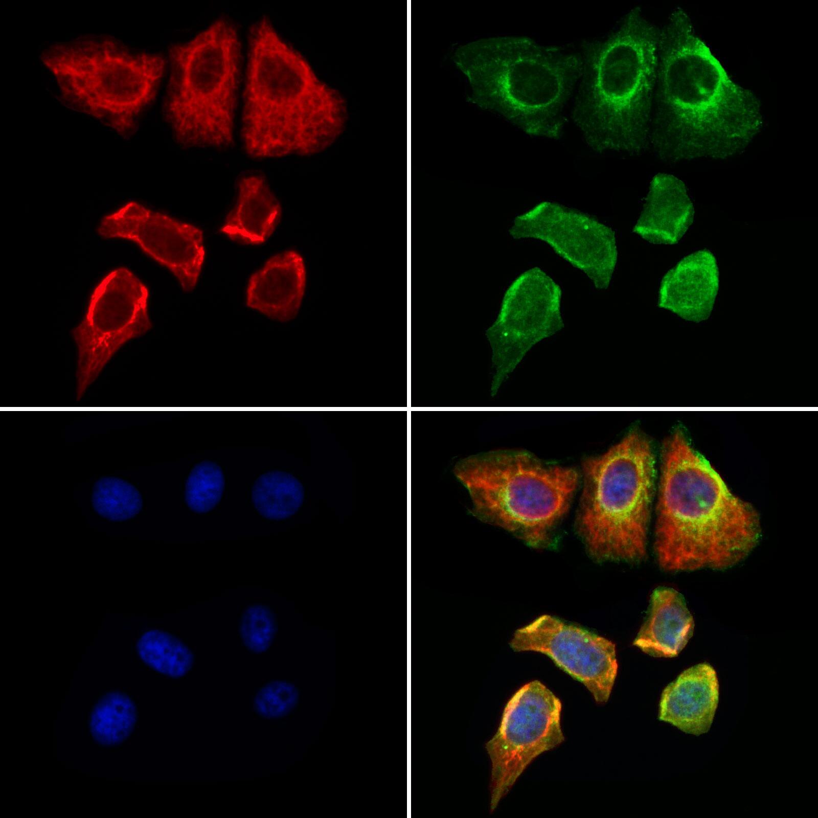
Rabbit anti-GPX4 Polyclonal Antibody staining Hela cells by IF/ICC. The samples were fixed with PFA and permeabilized in 0.1% Triton X-100,then blocked in 10% serum for 45 minutes at 25°C. Samples were then incubated with primary antibody(1:200) and mouse anti-beta tubulin antibody(1:200) for 1 hour at 37°C. An AlexaFluor594 conjugated goat anti-rabbit IgG(H+L) antibody(Red) and an AlexaFluor488 conjugated goat anti-mouse IgG(H+L) antibody(Green) were used as the secondary antibody.
The nuclear counter stain is DAPI(blue).

 危险品化学品经营许可证(不带存储) 许可证编号:沪(杨)应急管危经许[2022]202944(QY)
  营业执照(三证合一)