


 用小程序,查商品更便捷


Specificity/Sensitivity
Species Reactivity:
Human, Mouse
参考图片
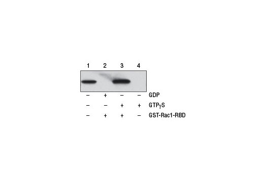
Figure 1. NIH/3T3 cell lysates (500 µl at 1 mg/ml) were treated in vitro with GTPγS or GDP to activate or inactivate Rac1 (refer to optional step C in protocol). The lysates were then incubated with glutathione resin and GST-PAK1-PBD (lanes 2 and 3). GTPγS-treated lysate was also incubated without GST-PAK1-PBD in the presence of glutathione resin as a negative control (lane 4). Western blot analysis of cell lysate (20 µg, lane 1) or 20 µl of the eluted samples (lanes 2, 3, and 4) was performed using a Rac1 Mouse mAb. Anti-mouse IgG, HRP-linked Antibody #7076 was used as the secondary antibody.图1. NIH/3T3细胞提取物(500 µl at 1 mg/ml)在体外使用GTPγS或 GDP处理以激活或失活Rac1(参考步骤C)。这些裂解液随后被谷胱甘肽树脂和GST-PAK1-PBD (lanes 2 和3)孵育。GTPγS处理的裂解液也在缺乏GST-PAK1-PBD而存在谷胱甘肽树脂作为阴性对照的情况下孵育(lane4)。使用Rac1 Mouse mAb对细胞裂解液(20 µg, lane 1)或20ul 稀释样品(lanes 2, 3, 和 4))进行western blot分析。Anti-mouse IgG, HRP-linked Antibody #7076作为二抗。
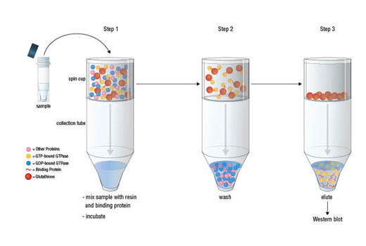
Figure 2. The GTP-bound GTPase pull-down process can be divided into 3 steps as shown. Step 1: Mix sample, binding protein, and glutathione resin in the spin cup and incubate at 4ºC to allow GTP-bound GTPase binding to the glutathione resin through GST-linked binding protein. Step 2: Remove unbound proteins by centrifugation. Step 3: Elute glutathione resin-bound GTPase with SDS buffer. The eluted sample can then be analyzed by western blot.图2.如图所示, GTP-bound GTPase pull-down过程可以被分成3个步骤。第一步:在自旋杯中混合样品,结合蛋白,和谷胱甘肽树脂,4ºC孵育以使GTP-bound GTPase与谷胱甘肽树脂通过GST- linked结合蛋白结合。第二步:通过离心去除没有结合的蛋白。第三步:使用SDS缓冲液稀释谷胱甘肽树脂。稀释的样品可以随后用于western blot分析。

 危险品化学品经营许可证(不带存储) 许可证编号:沪(杨)应急管危经许[2022]202944(QY)
  营业执照(三证合一)