


 用小程序,查商品更便捷


WB
1:1000IHC-P
1:250
Tyrosine-protein kinase transmembrane receptor ROR1, also known as neurotrophic tyrosine kinase, receptor-related 1 (NTRKR1), is an enzyme that in humans is encoded by the ROR1 gene. ROR1 is a member of the receptor tyrosine kinase-like orphan receptor (ROR) family. The protein encoded by this gene is a receptor tyrosine kinase that modulates neurite growth in the central nervous system. It is a type I membrane protein and belongs to the ROR subfamily of cell surface receptors. The expression of soluble ROR1 does not differ between normal individuals and tumor patients, and the expression level in serum is low or undetectable, independent of disease progression and severity. However, intact membrane bound ROR1 is specifically expressed at high levels in various tumor tissues. ROR1 is currently under investigation for its role in the metastasis of cancer cells. ROR1 has been found in tumor cells of many different types of cancer, such as chronic lymphocytic leukemia (CLL) and other hematological malignancies. It appears that ROR1 is mainly expressed in poorly differentiated tumors with high potential for recurrence and metastasis.
12 months from date of receipt / reconstitution, 4 °C as supplied
参考图片
WB result of ROR1 Rabbit mAb
Primary antibody: ROR1 Rabbit mAb at 1/1000 dilution
Lane 1: K-562 whole cell lysate 20 µg
Lane 2: MCF7 whole cell lysate 20 µg
Lane 3: PANC-1 whole cell lysate 20 µg
Lane 4: MDA-MB-231 whole cell lysate 20 µg
Lane 5: A549 whole cell lysate 20 µg
Negative control: K-562 whole cell lysate; MCF7 whole cell lysate
Secondary antibody: Goat Anti-Rabbit IgG, (H+L), HRP conjugated at 1/10000 dilution
Predicted MW: 104 kDa
Observed MW: 130, 135 kDa
(This blot was developed with high sensitivity substrate)
IHC shows positive staining in paraffin-embedded human ovarian carcinoma. Anti-ROR1 antibody was used at 1/250 dilution, followed by a HRP Polymer for Mouse & Rabbit IgG (ready to use). Counterstained with hematoxylin. Heat mediated antigen retrieval with Tris/EDTA buffer pH9.0 was performed before commencing with IHC staining protocol.
IHC shows positive staining in paraffin-embedded human gastric carcinoma. Anti-ROR1 antibody was used at 1/250 dilution, followed by a HRP Polymer for Mouse & Rabbit IgG (ready to use). Counterstained with hematoxylin. Heat mediated antigen retrieval with Tris/EDTA buffer pH9.0 was performed before commencing with IHC staining protocol.
IHC shows positive staining in paraffin-embedded human endometrial carcinoma. Anti-ROR1 antibody was used at 1/250 dilution, followed by a HRP Polymer for Mouse & Rabbit IgG (ready to use). Counterstained with hematoxylin. Heat mediated antigen retrieval with Tris/EDTA buffer pH9.0 was performed before commencing with IHC staining protocol.
IHC shows positive staining in paraffin-embedded human mantle lymphoma. Anti-ROR1 antibody was used at 1/250 dilution, followed by a HRP Polymer for Mouse & Rabbit IgG (ready to use). Counterstained with hematoxylin. Heat mediated antigen retrieval with Tris/EDTA buffer pH9.0 was performed before commencing with IHC staining protocol.
Negative control: IHC shows negative staining in paraffin-embedded human tonsil. Anti-ROR1 antibody was used at 1/250 dilution, followed by a HRP Polymer for Mouse & Rabbit IgG (ready to use). Counterstained with hematoxylin. Heat mediated antigen retrieval with Tris/EDTA buffer pH9.0 was performed before commencing with IHC staining protocol.
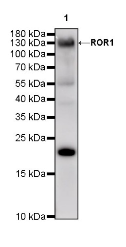
WB result of ROR1 Rabbit mAb
Primary antibody: ROR1 Rabbit mAb at 1/1000 dilution
Lane 1: NIH/3T3 whole cell lysate 20 µg
Secondary antibody: Goat Anti-Rabbit IgG, (H+L), HRP conjugated at 1/10000 dilution
Predicted MW: 104 kDa
Observed MW: 130 kDa
(This blot was developed with high sensitivity substrate)

 危险品化学品经营许可证(不带存储) 许可证编号:沪(杨)应急管危经许[2022]202944(QY)
  营业执照(三证合一)