


 全部商品分类
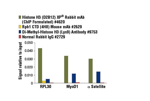


 用小程序,查商品更便捷


The SimpleChIP® Enzymatic Chromatin IP Kit (Agarose Beads) #9002 contains the buffers and reagents necessary to perform up to 6 chromatin preparations and 30 chromatin immunoprecipitations and is optimized for 4 X 10⁶cells per experiment. A complete assay can be performed in as little as two days and can easily be scaled up or down for use with more or fewer cells.
Cells are fixed with formaldehyde and lysed, and chromatin is fragmented by partial digestion with Micrococcal Nuclease to obtain chromatin fragments of 1 to 5 nucleosomes. Enzymatic fragmentation of chromatin is much milder than sonication and eliminates problems resulting from variability in sonication power and emulsification of chromatin during sonication, which can result in incomplete fragmentation of chromatin or loss of antibody epitopes due to protein denaturation and degradation. Chromatin immunoprecipitations are performed using ChIP-validated antibodies and ChIP-Grade Protein G Agarose Beads. After reversal of protein-DNA cross-links, the DNA is purified using DNA purification spin columns, allowing for easy and efficient recovery of DNA and removal of protein contaminants without the need for phenol/chloroform extractions and ethanol precipitations. The enrichment of particular DNA sequences during immunoprecipitation can be analyzed by a variety of methods, including standard PCR and quantitative real-time PCR. Please note that this kit is not compatible with ChIP-seq because the ChIP-Grade Protein G Agarose Beads are blocked with sonicated salmon sperm DNA, which interferes with downstream sequencing.
The SimpleChIP® Kit also provides important controls to ensure a successful ChIP experiment. The kit contains a positive control Histone H3 Antibody, a negative control Normal Rabbit IgG Antibody and primer sets for PCR detection of the human and mouse ribosomal protein L30 (RPL30) genes. Histone H3 is a core component of chromatin and is bound to most DNA sequences throughout the genome, including the RPL30 locus. Thus, the Histone H3 Antibody provides a universal positive control that should enrich for almost any locus examined.

Specificity/Sensitivity
Please store components at the temperatures indicated on the individual tube labels.
参考图片
FIGURE 3. Chromatin immunoprecipitations were performed using digested chromatin from HeLa cells and the indicated ChIP-validated antibodies. Purified DNA was analyzed by quantitative real-time PCR, using SimpleChIP® Human RPL30 Exon 3 Primers #7014 (control primer set), SimpleChIP® Human MyoD1 Exon 1 Primers #4490, and SimpleChIP® Human α Satellite Repeat Primers #4486. The amount of immunoprecipitated DNA in each sample is represented as signal relative to the total amount of input chromatin (equivalent to 1).
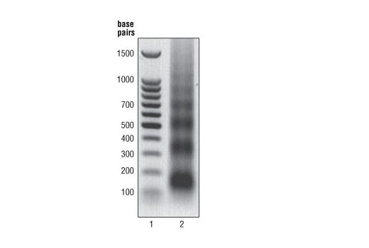
FIGURE 1. HeLa cells were formaldehyde-crosslinked and chromatin was prepared and digested as described in Section A of protocol. DNA was purified as described in Section B and 10 μl were separated by electrophoresis on a 1% agarose gel (lane 2) and stained with ethidium bromide. Lane 2 shows that the majority of chromatin was digested to 1 to 5 nucleosomes in length (150 to 900 bp).
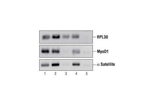
FIGURE 2. Chromatin immunoprecipitations were performed using digested chromatin from HeLa cells and either Histone H3 (D2B12) XP® Rabbit mAb (ChIP Formulated) #4620 (lane 2), Rpb1 CTD (4H8) Mouse mAb #2629 (lane 3), Di-Methyl Histone H3 (Lys9) Antibody #9753 (lane 4) or Normal Rabbit IgG #2729 (lane 5). Purified DNA was analyzed by standard PCR methods using SimpleChIP® Human RPL30 Exon 3 Primers #7014, SimpleChIP® Human MyoD1 Exon 1 Primers #4490, and SimpleChIP® Human α Satellite Repeat Primers #4486. PCR products were observed for each primer set in the input sample (lane 1) and various protein-specific immunoprecipitations but no PCR products were observed with immunoprecipitation using Normal Rabbit IgG #2729 (lane 5).