


 下载产品说明书
 用小程序,查商品更便捷


 收藏
收藏
 对比
对比 咨询
咨询
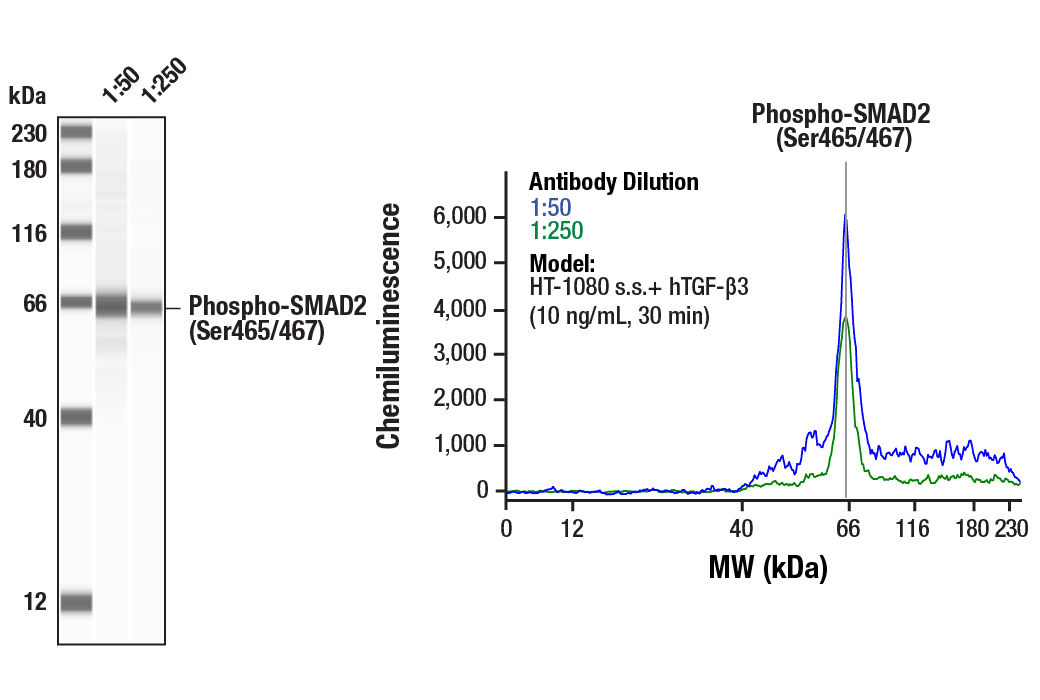
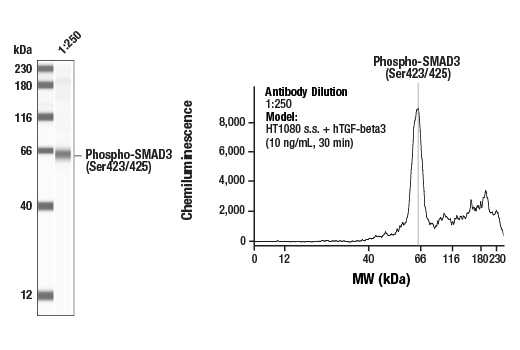
Specificity/Sensitivity
参考图片
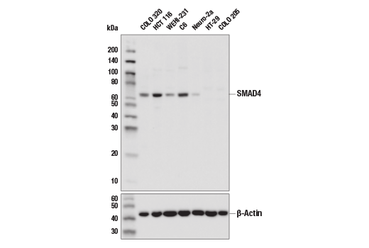
Western blot analysis of extracts from various cell lines using Smad4 (D3M6U) Rabbit mAb (upper) and β-Actin (D6A8) Rabbit mAb #8457 (lower). HT-29 and COLO 205 are Smad4-null mutant cell lines, confirming specificity of the antibody.
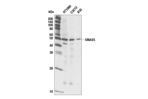
Western blot analysis of extracts from HT1080 (human), C2C12 (mouse) and B35 (rat) using Smad3 (C67H9) Rabbit mAb.
Western blot analysis of extracts from HT1080 cells, treated with TGF-β1, TGFR inhibitor SB-431542 or BMP-2, using Phospho-Smad3 (Ser423/425) (C25A9) Rabbit mAb #9520 (upper) or total Smad3 (C67H9) Rabbit mAb #9523 (lower).
Confocal immunofluorescent analysis of HT1080 cells, untreated (left) or TGFβ-treated (right), using Smad3 (C67H9) Rabbit mAb (green). Actin filaments have been labeled with Alexa Fluor® 555 phalloidin (red).
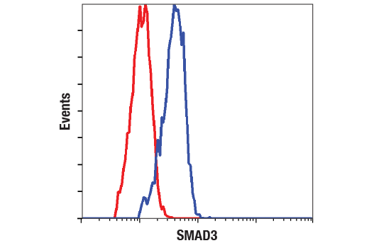
Flow cytometric analysis of HT-1080 cells using Smad3 (C67H9) Rabbit mAb #9523 (blue) compared to a nonspecific negative control antibody (red).
After the primary antibody is bound to the target protein, a complex with HRP-linked secondary antibody is formed. The LumiGLO* is added and emits light during enzyme catalyzed decomposition.
Western blot analysis of extracts from COS, NIH3T3, PC12, and SK-N-MC cells, using Smad4 Antibody.
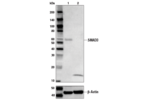
Western blot analysis of extracts from NIH/3T3 cells, transfected with 100 nM SignalSilence® Control siRNA (Unconjugated) #6568 (-) or SignalSilence® Smad4 siRNA I (Mouse Specific) (+), using Smad4 Antibody #9515 (upper) or β-Actin (D6A8) Rabbit mAb #8457 (lower). The Smad4 Antibody confirms silencing of Smad4 expression, while the β-Actin (D6A8) Rabbit mAb is used as a loading control.
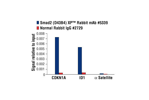
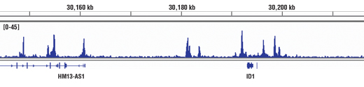
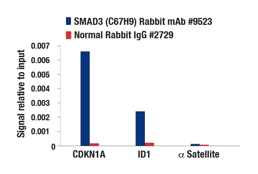
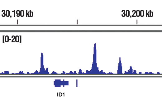
Chromatin immunoprecipitations were performed with cross-linked chromatin from 4 x 10⁶ HaCaT cells treated with Human TGF-β3 #3706 (7 ng/ml) for 1 h and either 10 μl of Smad3 (C67H9) Rabbit mAb or 2 μl of Normal Rabbit IgG #2729 using SimpleChIP® Enzymatic Chromatin IP Kit (Magnetic Beads) #9003. The enriched DNA was quantified by real-time PCR using SimpleChIP® Human CDKN1A Intron 1 Primers #4669, SimpleChIP® Human ID1 Promoter Primers #5139, and SimpleChIP® Human α Satellite Repeat Primers #4486. The amount of immunoprecipitated DNA in each sample is represented as signal relative to the total amount of input chromatin, which is equivalent to one.
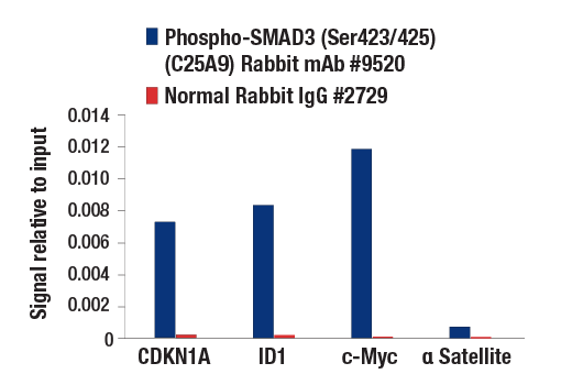
Chromatin immunoprecipitations were performed with cross-linked chromatin from 4 x 10⁶ HaCaT cells treated with Human TGF-β3 #3706 (7 ng/ml) for 1 h and either 5 μl of Phospho-Smad3 (Ser423/425) (C25A9) Rabbit mAb or 2 μl of Normal Rabbit IgG #2729 using SimpleChIP® Enzymatic Chromatin IP Kit (Magnetic Beads) #9003. The enriched DNA was quantified by real-time PCR using SimpleChIP® Human CDKN1A Intron 1 Primers #4669, SimpleChIP® Human ID1 Promoter Primers #5139, human c-Myc intron 1 primers, and SimpleChIP® Human α Satellite Repeat Primers #4486. The amount of immunoprecipitated DNA in each sample is represented as signal relative to the total amount of input chromatin, which is equivalent to one.
Chromatin immunoprecipitations were performed with cross-linked chromatin from 4 x 10⁶ HaCaT cells treated with Human TGF-β3 #3706 (7ng/ml) for 1 h and either 20 μl of Smad4 Antibody or 2 μl of Normal Rabbit IgG #2729 using SimpleChIP® Enzymatic Chromatin IP Kit (Magnetic Beads) #9003. The enriched DNA was quantified by real-time PCR using SimpleChIP® Human CDKN1A Intron 1 Primers #4669, SimpleChIP® Human ID1 Promoter Primers #5139, and SimpleChIP® Human α Satellite Repeat Primers #4486. The amount of immunoprecipitated DNA in each sample is represented as signal relative to the total amount of input chromatin, which is equivalent to one.
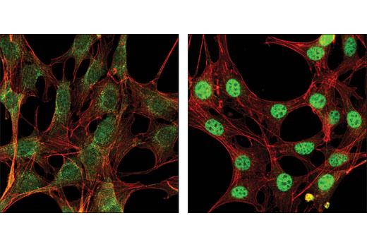
Confocal immunofluorescent analysis of NIH/3T3 cells, serum-starved (left) or treated with hTGF-β3 #8425 (right), using Smad2 (D43B4) XP® Rabbit mAb (green). Actin filaments have been labeled with DY-554 phalloidin (red).
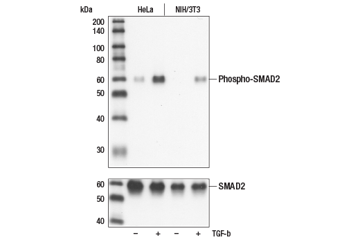
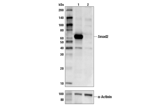
Western blot analysis of extracts from untreated or TGF-beta treated HeLa and NIH/3T3 cells, using Phospho-Smad2 (Ser465/467) (138D4) Rabbit mAb (upper), or Smad2 Antibody #3102 (lower).
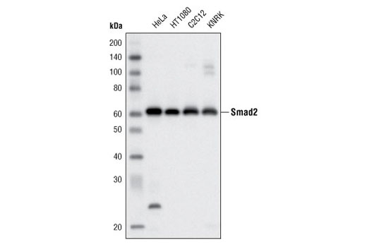
Western blot analysis of extracts from various cell lines using Smad2 (D43B4) XP® Rabbit mAb.
Chromatin immunoprecipitations were performed with cross-linked chromatin from 4 x 10⁶ HaCaT cells treated with Human TGF-β3 #8425 (7 ng/ml) for 1 h and either 10 μl of Smad2 (D43B4) XP® Rabbit mAb #5339 or 2 μl of Normal Rabbit IgG #2729 using SimpleChIP® Enzymatic Chromatin IP Kit (Magnetic Beads) #9003. The enriched DNA was quantified by real-time PCR using SimpleChIP® Human CDKN1A Intron 1 Primers #4669, SimpleChIP® Human ID1 Promoter Primers #5139, and SimpleChIP® Human α Satellite Repeat Primers #4486. The amount of immunoprecipitated DNA in each sample is represented as signal relative to the total amount of input chromatin, which is equivalent to one.
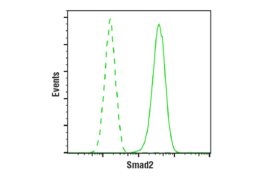
Flow cytometric analysis of HeLa cells using Smad2 (D43B4) XP® Rabbit mAb (blue) compared to a nonspecific negative control antibody (red).
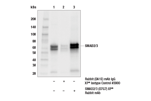
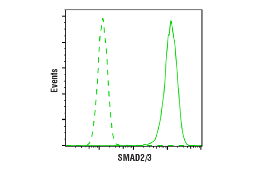
Flow cytometric analysis of HeLa cells using Smad2/3 (D7G7) XP® Rabbit mAb (blue) compared to Rabbit (DA1E) mAb IgG XP® Isotype Control #3900 (red).
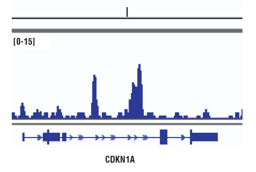
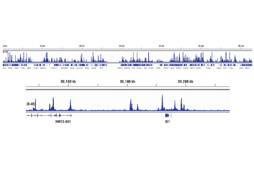
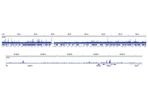
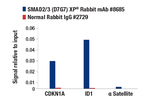
Chromatin immunoprecipitations were performed with cross-linked chromatin from 4 x 10⁶ HaCaT cells treated with hTGF-β3 #8425 (7 ng/ml, 1 hr) and either 10 μl of Smad2/3 (D7G7) XP® Rabbit mAb or 2 μl of Normal Rabbit IgG #2729 using SimpleChIP® Enzymatic Chromatin IP Kit (Magnetic Beads) #9003. The enriched DNA was quantified by real-time PCR using SimpleChIP® Human CDKN1A Intron 1 Primers #4669, SimpleChIP® Human ID1 Promoter Primers #5139, and SimpleChIP® Human α Satellite Repeat Primers #4486. The amount of immunoprecipitated DNA in each sample is represented as signal relative to the total amount of input chromatin, which is equivalent to one.
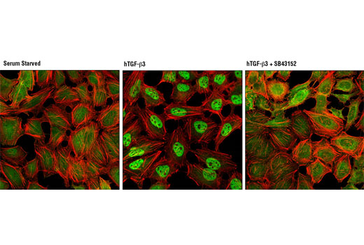
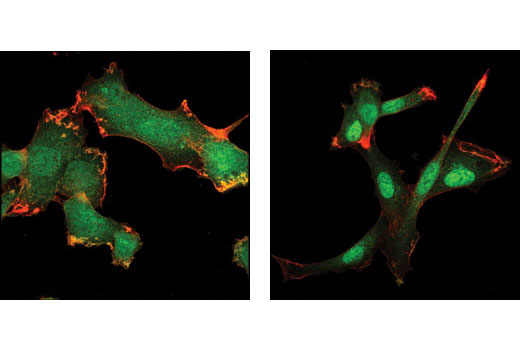
Confocal immunofluorescent analysis of HeLa cells, serum starved (left), treated with hTGF-β3 #8425 (100 ng/ml, 30 min, center), or treated with hTGF-β3 and SB43152 (10 ug/mL, 1 hr, right), using Smad2/3 (D7G7) XP® Rabbit mAb (green). Actin filaments were labeled with DY-554 phalloidin (red).
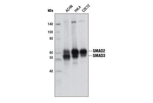
Western blot analysis of extracts from HeLa and ACHN cells using Smad2/3 (D7G7) XP® Rabbit mAb.
Western blot analysis of extracts from untreated (-) or TGF-β treated (+) HeLa and NIH/3T3 cells using Phospho-Smad2 (Ser465/467) (138D4) Rabbit mAb #3108 (upper) or Smad2 Antibody #3102 (lower).使用Phospho-Smad2 (Ser465/467) (138D4) Rabbit mAb #3108未处理(-)或Smad2抗体#3102(下)对未处理(-)或TGF-β treated (+)处理的HeLa细胞和NIH/3T3细胞提取物进行western blot分析。
Western blot analysis of extracts from various cell lines using Smad2 (D43B4) XP® Rabbit mAb #5339.使用Smad2 (D43B4) XP®Rabbit mAb#5339对多种细胞提取物进行western blot分析。
Western blot analysis of extracts from HeLa and ACHN cells using Smad2/3 (D7G7) XP® Rabbit mAb #8685.使用Smad2/3 (D7G7) XP®Rabbit mAb#8685对HeLa和ACHN细胞提取物进行western blot分析。
Western blot analysis of extracts from HT-1080 (human), C2C12 (mouse) and B35 (rat) cells using Smad3 (C67H9) Rabbit mAb #9523.使用Smad3 (C67H9)Rabbit mAb#9523对HT-1080 (human), C2C12 (mouse)和B35 (rat)提取物进行western blot分析。
Immunoprecipitation of Smad4 protein from HCT 116 cell extracts. Lane 1 is 10% input, lane 2 is Rabbit (DA1E) mAb IgG XP® Isotype Control #3900, and lane 3 is Smad4 (D3M6U) Rabbit mAb. Western blot analysis was performed using Smad4 (D3M6U) Rabbit mAb.
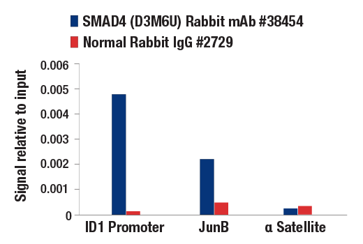
Chromatin immunoprecipitations were performed with cross-linked chromatin from 4 x 10⁶ HaCaT cells treated with TGF-β1 #8915 (7 ng/mL, 1 hr) and either 5 µl of Smad4 (D3M6U) Rabbit mAb or 2 µl of Normal Rabbit IgG #2729 using SimpleChIP® Enzymatic Chromatin IP Kit (Magnetic Beads) #9003. The enriched DNA was quantified by real-time PCR using SimpleChIP® Human ID1 Promoter Primers #5139, human JunB promoter primers, and SimpleChIP® Human α Satellite Repeat Primers #4486. The amount of immunoprecipitated DNA in each sample is represented as signal relative to the total amount of input chromatin (equivalent to one).
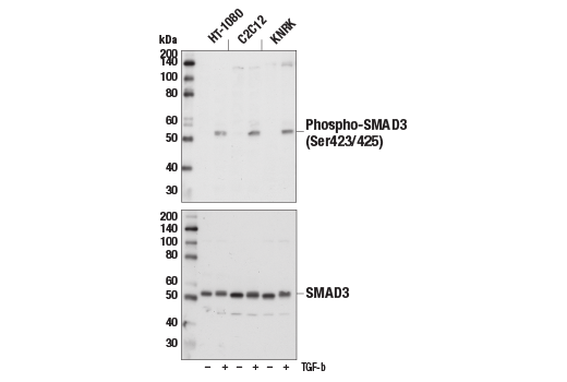
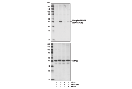
Western blot analysis of extracts from HT-1080, C2C12, or KNRK cells, untreated (-) or treated with TGF-β (10 ng/ml, 30 min; +), using Phospho-Smad3 (Ser423/425) (C25A9) Rabbit mAb (upper) or total Smad3 (C67H9) Rabbit mAb #9523 (lower).

 危险品化学品经营许可证(不带存储) 许可证编号:沪(杨)应急管危经许[2022]202944(QY)
  营业执照(三证合一)