


 下载产品说明书 下载SDS
 用小程序,查商品更便捷


 收藏
收藏
 对比
对比 咨询
咨询
参考图片
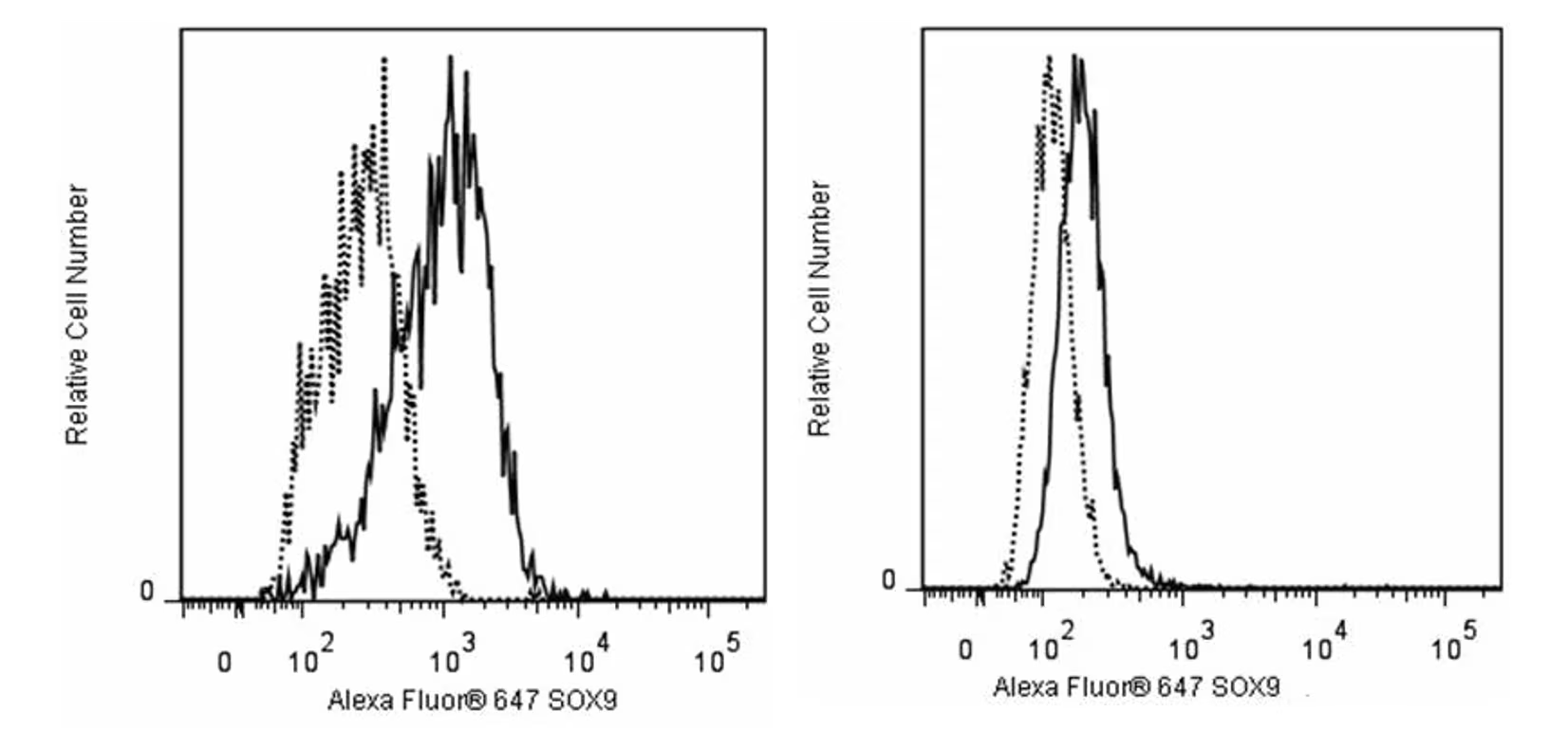
Flow cytometric analysis of SOX9 expression in human hepatocellular carcinoma and embryonic stem cell lines. Hep G2 cells (ATCC HB-8065, Left Panel) and H9 cells (WiCell, Madison, WI; Right Panel) were fixed with BD Cytofix™ Fixation Buffer (Cat. No. 554655), and permeabilized with BD Phosflow™ Perm Buffer III (Cat. No. 558050). The cells were stained with either Alexa Fluor® 647 Mouse IgG1 κ Isotype Control (Cat. No.557732; dashed line histogram) or Alexa Fluor® 647 Mouse Anti-Sox9 (Cat. No. 565493; solid line histogram). The fluorescence histograms were derived from gated events with the forward and side light-scatter characteristics of intact Hep G2 or H9 cells, respectively. Flow cytometric analysis was performed using a BD FACSCanto™ II Flow Cytometer System.
Flow cytometric analysis of SOX9 expression in human hepatocellular carcinoma and embryonic stem cell lines. Hep G2 cells (ATCC HB-8065, Left Panel) and H9 cells (WiCell, Madison, WI; Right Panel) were fixed with BD Cytofix™ Fixation Buffer (Cat. No. 554655), and permeabilized with BD Phosflow™ Perm Buffer III (Cat. No. 558050). The cells were stained with either Alexa Fluor® 647 Mouse IgG1 κ Isotype Control (Cat. No.557732; dashed line histogram) or Alexa Fluor® 647 Mouse Anti-Sox9 (Cat. No. 565493; solid line histogram). The fluorescence histograms were derived from gated events with the forward and side light-scatter characteristics of intact Hep G2 or H9 cells, respectively. Flow cytometric analysis was performed using a BD FACSCanto™ II Flow Cytometer System.

 危险品化学品经营许可证(不带存储) 许可证编号:沪(杨)应急管危经许[2022]202944(QY)
  营业执照(三证合一)