视频素材来源于JOVE https://www.jove.com/cn/v/58848/determination-regulatory-t-cell-subsets-murine-thymus-pancreatic
版权归原作者所有

△点击放大图片
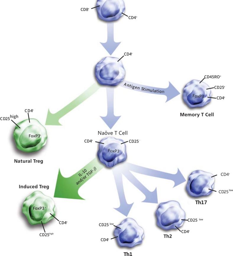
调节性T细胞的主要分类及特点
| 自然调节T细胞 | 适应性调节T细胞 | |||
| nTreg | Tr1 | Th3 | CD8+T | |
| 表型 | CD4+CD25int-hi, CD127low-dim | CD4+CD25- | CD4+CD25+from CD25- precursors | CD8+CD25+ |
| 其他相关的标志 | CTLA4+,GITR+, FoxP3+, CD127dim | CD25low-variable,CD45RBlow, FoxP3- | CD25low-variable, CD45RBlow, FoxP3+ | FoxP3+,CD28- |
| 发挥效应作用的机制 | 接触抑制;Granzyme B 依赖性,产生TGFβ | 通过细胞因子,产生IL-10 | 通过细胞因子,产生TGFβ | 接触抑制;通过细胞因子,产生IFN-γ、IL-6、IL-10 |
| 靶细胞 | APC & T effector cells | T effector cells | 未知 | APC & T effector cells |
| 体内的作用 | 抑制自身反应性T细胞 | 粘膜免疫及自身免疫反应 | 粘膜免疫及自身免疫反应 | 抑制自身反应性T细胞 |
| 体外扩增 | TCR/CD28共刺激及IL-2 | CD3, IL-10, Vitamin D | CD3, TGFβ | CD3 |
实验步骤
- 1
- 2
-
试剂准备
● 配制工作液之前,缓慢颠倒混匀5次以下试剂:BD Pharmingen™ TF Fix/Perm Buffer (4X), TF Diluent Buffer 以及 TF Perm/Wash Buffer (5X);
● 制备1x Fix/Perm 工作液:用TF Diluent Buffer3:1稀释 4x Fix/Perm Buffer 。每个样本通常需要1ml 1x Fix/Perm工作液。(如20个样本需5ml 4x Fix/Perm Buffer 和15ml TF Diluent Buffer)。1x Fix/Perm工作液配好后,尽量在1小时之内用完;
● 制备1x Perm/Wash 工作液:用去离子水4:1稀释5x Perm/Wash Buffer,每个样本需要7.5ml 1x Perm/Wash 工作液。如20个样本需30ml 5x Perm/Wash Buffer和120ml 去离子水 )。1x Perm/Wash Buffer可在 2-8°C保存一周;
● 胞内染色过程中所有工作液需置于冰上或 2-8°C。
buffer准备Fix/Permbuffer稀释 Perm/Wash buffer稀释,请参考上面的步
表面染色用100ul stain buffer制备单细胞悬液(10E6)CD4-FITC、CD25-PE抗体染色,2-8°C避光孵育20-30分钟设置空白管、补偿管、对照管、样本管
固定/破膜
振荡混匀细胞,加入1ml新鲜配制的1XFix/Permbuffer工作液,混匀2-8°度避光孵育40-50分钟
破膜/清洗
向已固定破膜的细胞中加入1XPerm/Wash buffer在2-8度条件下350g离心6分钟,去上清再加入2ml 1XPerm/Wash buffer,在2-8度条件下350g离心6分钟,去上清再加入2ml 1XPerm/Wash buffer,在2-8度条件下350g离心6分钟,去上清
核内染色
用80-100ul 的1XPerm/Wash buffer 重悬细胞加入FOXP3-APC标记抗体或非特异性对照,需涡旋振荡10s以充分混匀,2-8度避光孵育40-50分钟
破膜/清洗
清洗前轻轻振荡混匀样本加入2ml的1XPerm/Wash buffer,在2-8度条件下350g离心6分钟,去上清 -
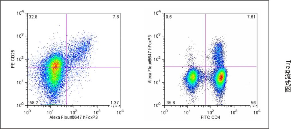
Human 外周血Treg染色案例:

△点击放大图片
Flow cytometric analysis of Alexa Fluor®Human PBMC were stained with FITC Anti-Human CD4 (clone RPA-T4, Cat. No. 555346) and PE Anti-Human CD25 (Clone M-A251, Cat. No. 555432)simultaneously. Cells were fixed and permeabilized (see recommended assay procedure) followed by intracellular staining with the AlexaFluor®based on light scattering characteristics of lymphocytes and fluorescence characteristics of CD4+ or CD25+ respectively,shown as either FoxP3 vs CD25 (left panel) or FoxP3 vs CD4 (right panel). Flow cytometry was performed on a BD FACSCalibur™ System.


