


 下载产品说明书
 用小程序,查商品更便捷


 收藏
收藏
 对比
对比 咨询
咨询
- 描述数量/尺寸零件号EntrezGene ID
- PE Rat Anti-Mouse CD4460 Tests (1 ea)51-9007324N/A
- PerCP-Cy™5.5 Rat Anti-Mouse CD460 Tests (1 ea)51-9007325N/A
- APC Rat Anti-Mouse CD62L60 Tests (1 ea)51-9007326N/A
- APC-Cy™7 Rat Anti-Mouse CD3 Molecular Complex60 Tests (1 ea)51-9007327N/A
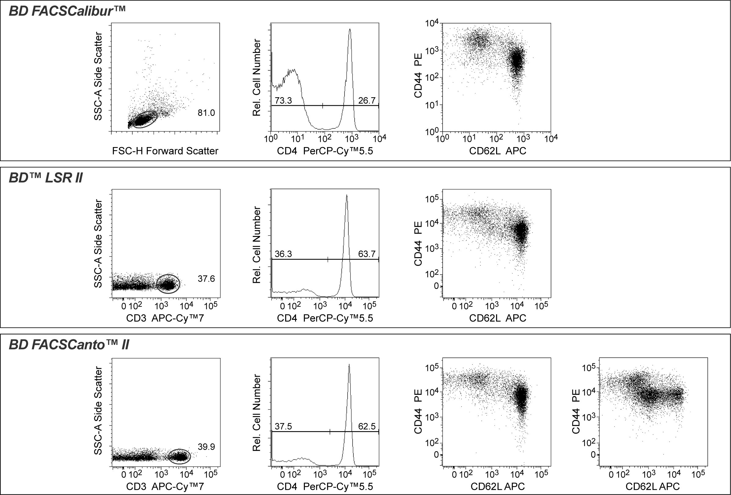
参考图片
Multicolor flow cytometric analysis of naïve and memory CD4+ T cell subsets defined by the coexpressed levels of CD62L and CD44. Splenocytes from a young (4 weeks) or older (9 weeks) BALB/c mouse donor were stained for analysis using the FACSCalibur™ System (Top Figures) with a combination (5 μl each) of PerCP-Cy™5.5 Rat Anti-Mouse CD4, PE Rat Anti-Mouse CD44, and APC Rat Anti-Mouse CD62L. Splenocytes were also stained with APC-Cy™7 Rat Anti-Mouse CD3 Molecular Complex, then analyzed on the BD™ LSR II and FACSCanto™ II systems (Middle and Bottom Figures, respectively). Two-color flow cytometric dot plots showing the expression of CD62L and CD44 (right dot plots) were derived from CD4+ cells (middle histograms) with the light scattering characteristics of viable lymphocytes (left FACSCalibur dotplot) or viable CD3+ cells (left LSR II and FACSCanto II dotplots). FSC vs SSC data from multiparameter analysis using the LSR II and FACSCanto II were also used for gating viable cells (data not shown). The patterns for the coexpressed levels of CD62L and CD44 by CD4+ T cells from the same young BALB/c mouse donor were very similar as determined by the 3 different flow cytometer systems (right dot plots). Interestingly, multicolor flow cytometric analysis of spleen cells from an older BALB/c mouse donor (far right dot plot from FACSCanto II analysis) revealed a significantly altered coexpression pattern of CD62L and CD44 by CD4+ T cells.

 危险品化学品经营许可证(不带存储) 许可证编号:沪(杨)应急管危经许[2022]202944(QY)
  营业执照(三证合一)