


 用小程序,查商品更便捷


Product Usage Information
| Application | Dilution |
|---|---|
| Western Blotting | 1:1000 |
| Immunoprecipitation | 1:50 |
| Immunofluorescence (Immunocytochemistry) | 1:50 - 1:100 |
| Flow Cytometry (Fixed/Permeabilized) | 1:50 - 1:200 |
Specificity/Sensitivity
物种反应性:
人, 小鼠
参考图片
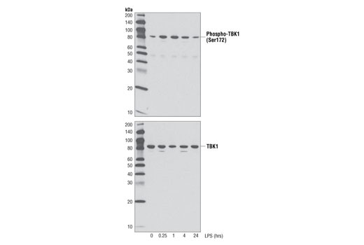
Western blot analysis of extracts from THP-1 cells differentiated with TPA #4174 (80 nM, overnight) followed by treatment with LPS (1 μg/ml), up to 24h, using Phospho-TBK1 (Ser172) (D52C2) XP® Rabbit mAb (upper), or total TBK1/NAK (D1B4) Rabbit mAb #3504 (lower).Western blot analysis of extracts from THP-1 cells differentiated with TPA #4174 (80 nM, overnight) followed by treatment with LPS (1 μg/ml), up to 24h, using Phospho-TBK1 (Ser172) (D52C2) XP® Rabbit mAb (upper), or total TBK1/NAK (D1B4) Rabbit mAb #3504 (lower).Western免疫印迹分析THP-1细胞的细胞抽提液,THP-1细胞经过 TPA #4174 (80 nM, 过夜)分化然后用LPS (1 μg/ml)处理多达24小时,所用抗体为Phospho-TBK1 (Ser172) (D52C2) XP® Rabbit mAb (上图)或total TBK1/NAK (D1B4) Rabbit mAb #3504 (下图)。
Flow cytometric analysis of THP-1 cells differentiated with TPA #9905, untreated (blue) or LPS-treated (green), using Phospho-TBK1/NAK (Ser172) (D52C2) XP® Rabbit mAb.流式细胞仪分析经过TPA #9905处理的THP-1细胞,未经其他处理的 (蓝色) 或 LPS处理的(绿色), 所用抗体为Phospho-TBK1/NAK (Ser172) (D52C2) XP® Rabbit mAb。
Confocal immunofluorescent analysis of THP-1 cells differentiated with TPA #4174 (80nM, overnight) (left), followed by treatment with LPS (1μg/ml, 1 hour) (center) or LPS with λ phosphatase treatment (right) using Phospho-TBK1/NAK (Ser172) (D52C2) XP® Rabbit mAb (green). Actin filaments were labeled with DY-554 Phalloidin (red). Blue pseudocolor = DRAQ5® #4084 (fluorescent DNA dye).共聚焦免疫荧光分析经不同处理的THP-1细胞,TPA #4174 (80nM,过夜) (左图), 经TPA处理后又经LPS (1μg/ml, 1小时)(中间图) 或 LPS 与λ磷酸酶处理(右图),所用抗体为Phospho-TBK1/NAK (Ser172) (D52C2) XP® Rabbit mAb (绿色)。肌动蛋白丝是用DY-554 Phalloidin 标记(红色)。Blue pseudocolor = DRAQ5® #4084 (DNA荧光染料)。
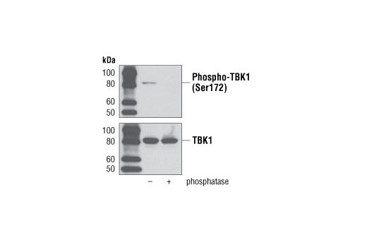
Western blot analysis of extracts from THP-1 cells differentiated with TPA #4174 (80 nM, overnight) followed by treatment with LPS (1 μg/ml, 1 hour), with or without phosphatase treatment using Phospho-TBK1 (Ser172) (D52C2) XP® Rabbit mAb (upper), or total TBK1/NAK (D1B4) Rabbit mAb #3504 (lower).Western免疫印迹分析THP-1细胞的细胞抽提液,THP-1细胞经过 TPA #4174 (80 nM, 过夜)分化然后用在磷酸酶存在或不存在下用LPS (1 μg/ml, 1小时)处理 所用抗体为Phospho-TBK1 (Ser172) (D52C2) XP® Rabbit mAb (上图) 或 total TBK1/NAK (D1B4) Rabbit mAb #3504 (下图)。

 危险品化学品经营许可证(不带存储) 许可证编号:沪(杨)应急管危经许[2022]202944(QY)
  营业执照(三证合一)