


 用小程序,查商品更便捷


Scientific Data
 View Larger
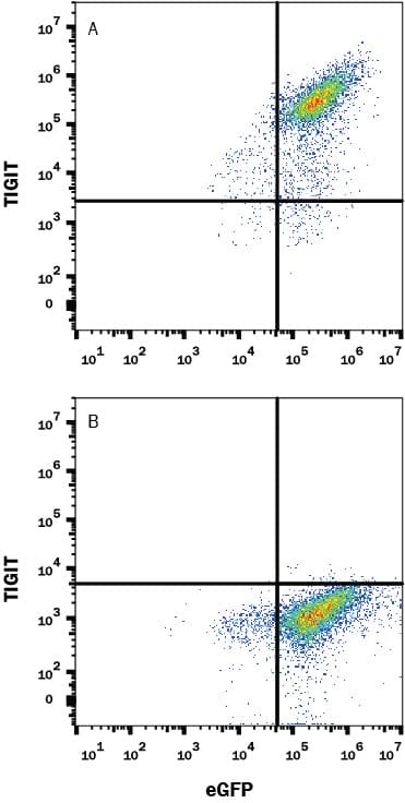
Detection of TIGIT in HEK293 Human Cell Line transfected with Cynomolgus Monkey TIGIT and eGFP by Flow Cytometry. HEK293 human embryonic kidney cell line transfected with either (A) cynomolgus monkey TIGIT or (B) irrelevant protein, and eGFP, was stained with Rabbit anti-cynomolgus monkey TIGIT monoclonal antibody (Catalog # MAB10532) followed by Allophycocyanin-conjugated anti-Rabbit IgG Secondary Antibody (F0111). Quadrant markers were set based on control antibody staining (MAB1050, data not shown). Staining was performed using our Staining Membrane-Associated Proteins protocol.
 View Larger
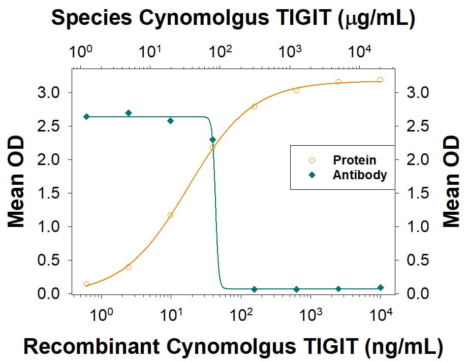
TIGIT binding to CD155/PVR blocked by TIGIT Antibody. In a functional ELISA, Recombinant Cynomolgus Monkey TIGIT (9380-TG) binds to immobilized Recombinant Human CD155/PVR (2530-CD) coated at 2.5 µg/mL (100 µL/well) in a dose-dependent manner (orange line). Binding is blocked (green line) by increasing concentrations of Rabbit Anti-Cynomolgus Monkey TIGIT Monoclonal Antibody (Catalog # MAB10532). At 70-350 ng/mL, this antibody will block 50% of the binding.
Cynomolgus Monkey TIGIT Antibody Summary
Met22-Pro142
Accession # XP_00554815
Applications
Please Note: Optimal dilutions should be determined by each laboratory for each application. General Protocols are available in the Technical Information section on our website.
Background: TIGIT
TIGIT (T cell Immunoreceptor with Ig and ITIM domains), also called VSTM3 (V-set and transmembrane domain-containing 3), VSIG9 (V-set and Ig domain-containing 9) and WUCAM (Washington University cell adhesion molecule) is a 30-34 kDa type I transmembrane protein that is a member of the CD28 family within the Ig superfamily of proteins (1-4). Human TIGIT cDNA encodes 244 amino acids (aa) including a 21 aa signal sequence, a 120 aa extracellular region with a V-type Ig-like domain and two potential N-glycosylation site, a 21 aa transmembrane sequence, and an 82 aa cytoplasmic domain with an ITIM motif (5). A 170 aa variant diverges after aa 166 (5). Within the ECD, human TIGIT shares only 68-75% aa sequence identity with mouse, porcine, canine, equine and bovine TIGIT. Cyno TIGIT shares 88.4% homology with human TIGIT. TIGIT is expressed on NK cells and subsets of activated, memory and regulatory T cells, and particularly on follicular helper T cells within secondary lymphoid organs (1, 2, 6-8). It binds to CD155/PVR/Necl-5 and Nectin-2/CD112/PVRL2 that appear on dendritic cells (DC) and endothelium (1-3, 7). Binding of TIGIT by DC induces IL-10 release and inhibits IL-12 production (2). Ligation of TIGIT on T cells down‑regulates TCR-mediated activation and subsequent proliferation, while NK cell TIGIT ligation blocks NK cell cytotoxicity (6-8). Through CD155 and Nectin-2, which also interact with DNAM-1/CD226 and CD96/Tactile, TIGIT is part of an interacting network of Ig superfamily members that may augment or oppose each other (3, 4, 6, 7). In particular, TIGIT binding to CD155 can antagonize the effects of DNAM-1 (6, 7). Soluble TIGIT is able to compete with DNAM-1 for CD155 binding and attenuates T cell responses, while mice lacking TIGIT show increased T cell responses and susceptibility to autoimmune challenges (2, 3, 8).
- Boles, K.S. et al. (2009) Eur. J. Immunol. 39:695.
- Yu, X. et al. (2009) Nat. Immunol. 10:48.
- Levin, S.D. et al. (2011) Eur. J. Immunol. 41:902.
- Xu, Z. et al. (2010) Cell. Mol. Immunol. 7:11.
- SwissProt Accession # Q495A1.
- Seth, S. et al. (2009) Eur. J. Immunol. 39:3160.
- Stanietsky, N. et al. (2009) Proc. Natl. Acad. Sci. USA 106:17858.
- Joller, N. et al. (2011) J. Immunol. 83:1338.
Preparation and Storage
- 12 months from date of receipt, -20 to -70 °C as supplied.
- 1 month, 2 to 8 °C under sterile conditions after reconstitution.
- 6 months, -20 to -70 °C under sterile conditions after reconstitution.

 危险品化学品经营许可证(不带存储) 许可证编号:沪(杨)应急管危经许[2022]202944(QY)
  营业执照(三证合一)