


 下载产品说明书 下载SDS
 用小程序,查商品更便捷


 收藏
收藏
 对比
对比 咨询
咨询

参考图片
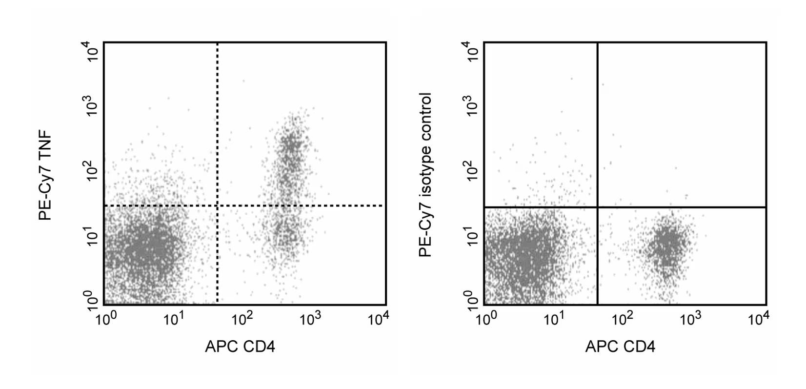
Expression of TNF by stimulated CD4+ and CD4- BALB/c spleen cells. Splenocytes from BALB/c mice were stimulated for 4 hours with PMA (5 ng/ml, Sigma, P-8139) and Ionomycin (500 ng, Sigma I-0634) in the presence of Brefeldin A (GolgiPlug, Cat. No. 555029). Cells were harvested, fixed, permeabilized and stained with APC-conjugated rat anti-mouse CD4 (APC-RM4-5, Cat. No. 553051) and either rat anti-mouse TNF antibody (PE-Cy7-MP6-XT22, Cat. No. 557644) (left panel) or immunoglobulin isotype control (PE-Cy7-R3-34, Cat. No. 557645), (right panel) by using BD Pharmingen staining protocol. To demonstrate specificity of staining the binding of PE-Cy7-MP6-XT22 was blocked by the preincubation of the conjugated antibody with molar excess of recombinant mouse TNF (0.25 µg, Cat. No. 554589, data not shown) and by preincubation of the fixed/permeabilized cells with an excess of unlabelled MP6-XT22 antibody (5 µg, Cat. No. 554416, data not shown) prior to staining. The quadrant markers for the bivariate dot plots were set based on the autofluorescence and isotype controls.
Expression of TNF by stimulated CD4+ and CD4- BALB/c spleen cells. Splenocytes from BALB/c mice were stimulated for 4 hours with PMA (5 ng/ml, Sigma, P-8139) and Ionomycin (500 ng, Sigma I-0634) in the presence of Brefeldin A (GolgiPlug, Cat. No. 555029). Cells were harvested, fixed, permeabilized and stained with APC-conjugated rat anti-mouse CD4 (APC-RM4-5, Cat. No. 553051) and either rat anti-mouse TNF antibody (PE-Cy7-MP6-XT22, Cat. No. 557644) (left panel) or immunoglobulin isotype control (PE-Cy7-R3-34, Cat. No. 557645), (right panel) by using BD Pharmingen staining protocol. To demonstrate specificity of staining the binding of PE-Cy7-MP6-XT22 was blocked by the preincubation of the conjugated antibody with molar excess of recombinant mouse TNF (0.25 µg, Cat. No. 554589, data not shown) and by preincubation of the fixed/permeabilized cells with an excess of unlabelled MP6-XT22 antibody (5 µg, Cat. No. 554416, data not shown) prior to staining. The quadrant markers for the bivariate dot plots were set based on the autofluorescence and isotype controls.

 危险品化学品经营许可证(不带存储) 许可证编号:沪(杨)应急管危经许[2022]202944(QY)
  营业执照(三证合一)