


 下载产品说明书 下载SDS
 用小程序,查商品更便捷


 收藏
收藏
 对比
对比 咨询
咨询Product Summary
Recovery
The recovery of mouse VEGF spiked to three levels throughout the range of the assay in various matrices was evaluated.
| Sample Type | Average % Recovery | Range % |
|---|---|---|
| Cell Culture Supernates (n=9) | 103 | 95-114 |
| EDTA Plasma (n=5) | 91 | 82-103 |
| Heparin Plasma (n=4) | 89 | 87-94 |
| Serum (n=9) | 101 | 83-115 |
| Tissue Homogenates (n=3) | 91 | 76-107 |
Linearity

Scientific Data

 View Larger
Mouse VEGF Quantikine ELISA Kit Increased production of proinflammatory cytokines and VEGF in the plasma and chondrocytes in high fat diet (HFD)-feeding mice.(a~d) levels of plasma IL-6 (a), TNF-alpha (b), IL-1 beta (c) and VEGF (d) in the C57BL/6 male mice fed with a normal diet (ND) or HFD for 8 weeks. (n = 5, **P < 0.01). (e~h) Relative mRNA expression of IL-6 (e), TNF-alpha (f), IL-1 beta (g) and VEGF (h) in the chondrocytes isolated from the 8-week ND or HFD-feeding mice. (n = 5, **P < 0.01). Image collected and cropped by CiteAb from the following open publication (https://pubmed.ncbi.nlm.nih.gov/26271607), licensed under a CC-BY license. Not internally tested by R&D Systems.
Assay Procedure
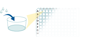
Refer to the product- Prepare all reagents, standard dilutions, and samples as directed in the product insert.
- Remove excess microplate strips from the plate frame, return them to the foil pouch containing the desiccant pack, and reseal.
- Add 50 µL of Assay Diluent to each well.
- Add 50 µL of Standard, Control, or sample to each well. Cover with a plate sealer, and incubate at room temperature for 2 hours.
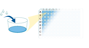
- Aspirate each well and wash, repeating the process 4 times for a total of 5 washes.
- Add 100 µL of Conjugate to each well. Cover with a new plate sealer, and incubate at room temperature for 2 hours.
- Aspirate and wash 5 times.
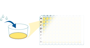
- Add 100 µL Substrate Solution to each well. Incubate at room temperature for 30 minutes. PROTECT FROM LIGHT.
- Add 100 µL of Stop Solution to each well. Read at 450 nm within 30 minutes. Set wavelength correction to 540 nm or 570 nm.
Mouse VEGF Quantikine ELISA Kit Summary

Background: VEGF
Vascular endothelial growth factor (VEGF or VEGF-A), also known as vascular permeability factor (VPF), is a potent mediator of both angiogenesis and vasculogenesis in the fetus and in adults (1-3). It is a member of the PDGF family that is characterized by the presence of eight conserved cysteine residues in a cystine knot structure and the formation of anti-parallel disulfide-linked dimers (4). Alternately spliced isoforms of 120, 164 and 188 amino acids (aa) have been found in mice, while 121, 145, 165, 183, 189, and 206 aa isoforms have been identified in humans (2, 4). In humans, VEGF165 appears to be the most abundant and potent isoform, followed by VEGF121 and VEGF189 (3, 4). The same pattern may exist in mice. Isoforms other than VEGF120 and VEGF121 contain basic heparin-binding regions and are not freely diffusible (4). Mouse VEGF164 shares 97% aa sequence identity with corresponding regions of rat VEGF. It also shares 89% aa sequence identity with human and porcine VEGF, 88% with bovine VEGF, and 90% with feline, equine, and canine VEGF. VEGF is expressed in multiple cells and tissues including skeletal and cardiac muscle (5, 6), hepatocytes (7), osteoblasts (8), neutrophils (9), macrophages (10), keratinocytes (11), brown adipose tissue (12), CD34+ stem cells (13), endothelial cells (14), fibroblasts, and vascular smooth muscle cells (15). VEGF expression is induced by hypoxia and cytokines such as IL-1, IL-6, IL-8, Oncostatin M, and TNF-alpha (3, 4, 9, 16). The isoforms are differentially expressed during development and in the adult (3).

 危险品化学品经营许可证(不带存储) 许可证编号:沪(杨)应急管危经许[2022]202944(QY)
  营业执照(三证合一)